 Explore
 Validate
 Learn
Learn Western blot
 ELISA
ELISA Immunocytochemistry
Antibody data
- Antibody Data
- Antigen structure
- References [0]
- Comments [0]
- Validations
- Western blot [1]
- Immunocytochemistry [2]
- Immunohistochemistry [6]
Submit
Validation data
Reference
Comment
Report error
- Product number
- LS-C669062 - Provider product page

- Provider
- LSBio
- Product name
- PDCD1 / CD279 / PD-1 Antibody (clone 7H6) LS-C669062
- Antibody type
- Monoclonal
- Description
- Protein A purified
- Reactivity
- Human
- Host
- Mouse
- Isotype
- IgG
- Antibody clone number
- 7H6
- Storage
- Store at 4°C for up 3 months. Store at -20°C for up to 1 year. Avoid freeze/thaw cycles.
No comments: Submit comment
Enhanced validation
- Submitted by
- LSBio (provider)
- Enhanced method
- Genetic validation
- Main image
- Experimental details
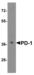
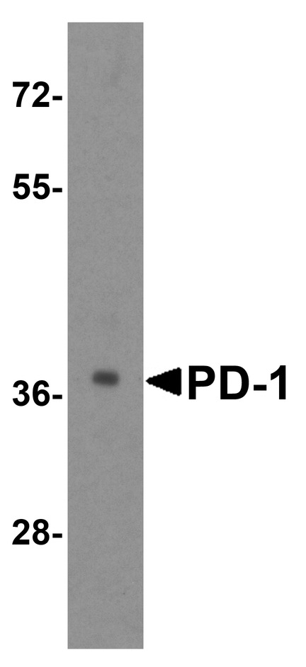
- Western blot analysis of PD-1 in transfected 293 cell lysate with PD-1 antibody at 1 ug/mL.
Supportive validation
- Submitted by
- LSBio (provider)
- Enhanced method
- Genetic validation
- Main image

- Experimental details
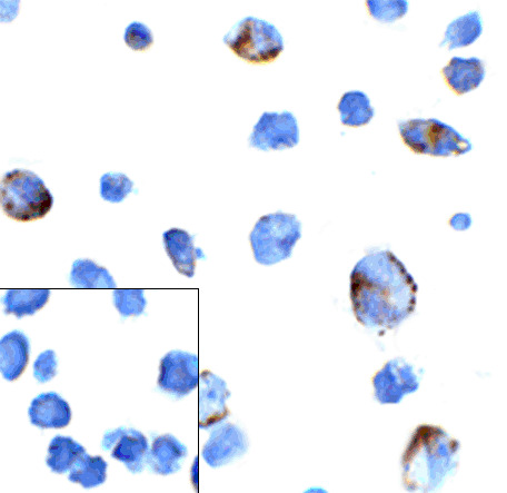
- Immunocytochemistry of PD-1 in transfected 293 cells with PD-1 antibody at 5 ug/mL. Lower left: Immunocytochemistry in transfected 293 cells with control mouse IgG antibody at 5 ug/mL.
- Submitted by
- LSBio (provider)
- Main image

- Experimental details
- Immunocytochemistry of PD-1 in transfected 293 cells with PD-1 antibody at 5 ug/mL. Lower left: Immunocytochemistry in transfected 293 cells with control mouse IgG antibody at 5 ug/mL.

Supportive validation
- Submitted by
- LSBio (provider)
- Enhanced method
- Genetic validation
- Main image
- Experimental details
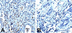
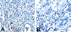
- Immunohistochemistry of PD-1 in (A) human breast cancer tissue and (B) human normal breast tissue with PD-1 antibody at 5 ug/mL.

- Submitted by
- LSBio (provider)
- Enhanced method
- Genetic validation
- Main image

- Experimental details
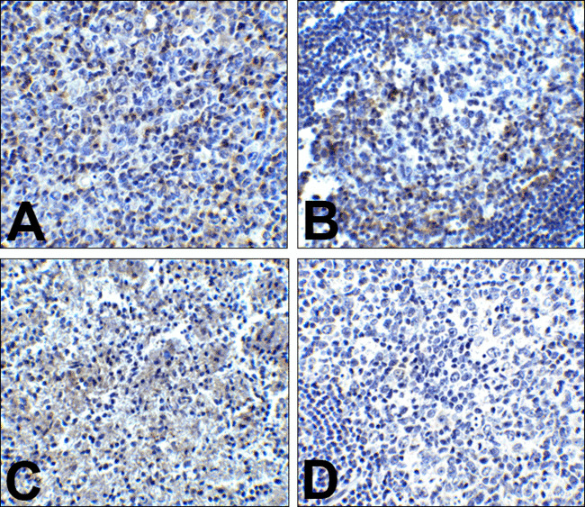
- Immunohistochemistry of PD-1 in (A) human tonsil tissue, (B) human lymph node tissue, and (C) human spleen tissue with PD-1 antibody at 5 ug/mL. (D) Immunohistochemistry in human tonsil tissue with control mouse IgG staining at 5 ug/mL.

- Submitted by
- LSBio (provider)
- Enhanced method
- Genetic validation
- Main image
- Experimental details
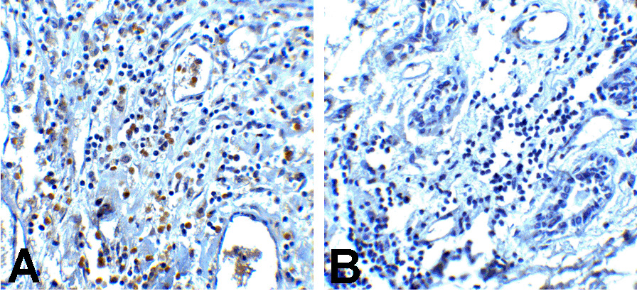
- Immunohistochemistry of PD-1 in (A) human breast cancer tissue and (B) human normal breast tissue with PD-1 antibody at 5 ug/mL.
- Submitted by
- LSBio (provider)
- Enhanced method
- Genetic validation
- Main image

- Experimental details
- Immunohistochemistry of PD-1 in (A) human tonsil tissue, (B) human lymph node tissue, and (C) human spleen tissue with PD-1 antibody at 5 ug/mL. (D) Immunohistochemistry in human tonsil tissue with control mouse IgG staining at 5 ug/mL.

- Submitted by
- LSBio (provider)
- Enhanced method
- Genetic validation
- Main image

- Experimental details
- Immunohistochemistry of PD-1 in (A) human breast cancer tissue and (B) human normal breast tissue with PD-1 antibody at 5 ug/mL.
