Explore
 Validate
 Learn
Learn Western blot
Antibody data
- Antibody Data
- Antigen structure
- References [0]
- Comments [0]
- Validations
- Western blot [1]
- Flow cytometry [2]
Submit
Validation data
Reference
Comment
Report error
- Product number
- 10-2001-25 - Provider product page

- Provider
- ABEOMICS Inc.
- Product name
- Recombinant Human PD-1 rabbit monoclonal Antibody Antibody
- Antibody type
- Monoclonal
- Description
- PDCD-1 (programmed cell death-1 protein), also designated CD279, is a type I transmembrane receptor and a member of the immunoglobin gene superfamily. It is expressed on activated T-cells, B-cells, and myeloid cells. Anti-PDCD-1 is a marker of angioimmunoblastic lymphoma and suggests a unique cell of origin for this neoplasm. Unlike CD10 and BCL6, PDCD-1 is expressed by few B-cells, so anti-PDCD-1 may be a more specific and useful diagnostic marker in angioimmunoblastic lymphoma. In addition, PDCD-1 expression provides evidence that angioimmunoblastic lymphoma is a neoplasm derived from germinal center-associated T-cells.
- Reactivity
- Human
- Conjugate
- Unconjugated
- Isotype
- IgG
- Antibody clone number
- ABMRR01
- Vial size
- 100 µg
- Concentration
- 0.5 mg/ml
- Storage
- Store the antibody at 4°C, stable for 6 months. For long-term storage, store at -20°C. Avoid repeat freez thawing
No comments: Submit comment
Supportive validation
- Submitted by
- ABEOMICS Inc. (provider)
- Main image
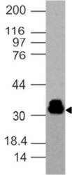
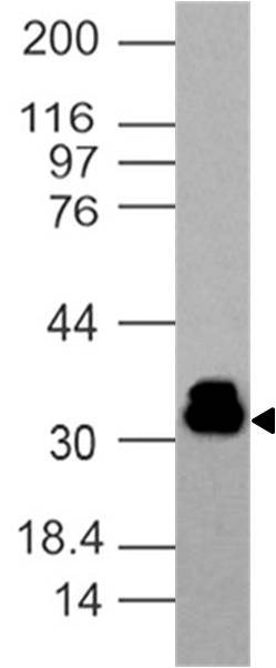
- Experimental details
- Western blot analysis of Rabbit recombinant hPD-1 antibody. Anti-Rabbit recombinant hPD-1 antibody was tested at 1 µg/ml in Jurkat cell lysate
- Protocol
- Protocol
Supportive validation
- Submitted by
- ABEOMICS Inc. (provider)
- Main image
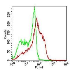
- Experimental details
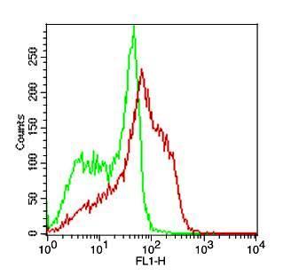
- Cell surface flow analysis of Rabbit recombinant hPD-1 antibody in PHA treated PBMC (Lymphocyte gated) using 0.5 µg/10^6 cells of Rabbit recombinant hPD-1 antibody (Clone:ABMRR01). Green represent isotype control and red represent Rabbit recombinant hPD-1 antibody. Goat anti rabbit FITC conjugate was used as secondary antibody.
- Protocol
- Protocol
- Submitted by
- ABEOMICS Inc. (provider)
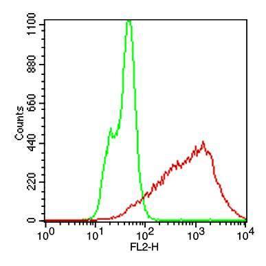
- Main image
- Experimental details
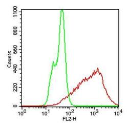
- Cell surface flow analysis of Rabbit recombinant h PD-1 antibody in CHO-PD1 transfected cell line using 0.2 µg/10^6 cells of Rabbit recombinant hPD-1 antibody (Clone: ABMRR01). Green represent CHO/K1 cells and red represent Rabbit recombinant hPD-1 antibody. Goat anti rabbit PE conjugate was used as secondary antibody.
- Protocol
- Protocol