 Explore
 Validate
 Learn
Learn Western blot
Antibody data
- Antibody Data
- Antigen structure
- References [2]
- Comments [0]
- Validations
- Western blot [6]
- Immunohistochemistry [3]
- Other assay [2]
Submit
Validation data
Reference
Comment
Report error
- Product number
- PA5-20350 - Provider product page

- Provider
- Invitrogen Antibodies
- Product name
- PD-1 Polyclonal Antibody
- Antibody type
- Polyclonal
- Antigen
- Synthetic peptide
- Description
- Despite its predicted molecular weight, PD-1 often migrates at a higher molecular weight in SDS-PAGE. A suggested positive control is THP-1 cell lysate.
- Concentration
- 1 mg/mL
Submitted references In PD-1+ human colon cancer cells NIVOLUMAB promotes survival and could protect tumor cells from conventional therapies.
Targeting CXCR4 potentiates anti-PD-1 efficacy modifying the tumor microenvironment and inhibiting neoplastic PD-1.
Ieranò C, Righelli D, D'Alterio C, Napolitano M, Portella L, Rea G, Auletta F, Santagata S, Trotta AM, Guardascione G, Liotti F, Prevete N, Maiolino P, Luciano A, Barbieri A, Di Mauro A, Roma C, Esposito Abate R, Tatangelo F, Pacelli R, Normanno N, Melillo RM, Scala S
Journal for immunotherapy of cancer 2022 Mar;10(3)
Journal for immunotherapy of cancer 2022 Mar;10(3)
Targeting CXCR4 potentiates anti-PD-1 efficacy modifying the tumor microenvironment and inhibiting neoplastic PD-1.
D'Alterio C, Buoncervello M, Ieranò C, Napolitano M, Portella L, Rea G, Barbieri A, Luciano A, Scognamiglio G, Tatangelo F, Anniciello AM, Monaco M, Cavalcanti E, Maiolino P, Romagnoli G, Arra C, Botti G, Gabriele L, Scala S
Journal of experimental & clinical cancer research : CR 2019 Oct 28;38(1):432
Journal of experimental & clinical cancer research : CR 2019 Oct 28;38(1):432
No comments: Submit comment
Supportive validation
- Submitted by
- Invitrogen Antibodies (provider)
- Main image

- Experimental details
- Western blot analysis of THP-1 cell lysate using a CD279/PD1 polyclonal antibody (Product # PA5-20350) in the (A) absence or (B) presence of blocking peptide at a 1 µg/mL dilution.

- Submitted by
- Invitrogen Antibodies (provider)
- Main image

- Experimental details
- Western Blot analysis of PD-1 in HeLa WT or PD-1 KD cells. Lysates (10 µg) were loaded onto SDS-PAGE and blots were probed with PD-1 Polyclonal Antibody (Product # PA5-20350) diluted to 4 µg/mL and anti-beta actin diluted to 1 µg/mL. 1 h incubation at RT in 0.05 NFDM/TBST. Secondary: Goat Anti-Rabbit IgG HRP conjugate at 1:10,000 dilution.

- Submitted by
- Invitrogen Antibodies (provider)
- Main image

- Experimental details
- Western Blot analysis of PD-1 in HeLa WT or PD-1 KO cells. Lysates (10 µg) were loaded onto SDS-PAGE and blots were probed with PD-1 Polyclonal Antibody (Product # PA5-20350) diluted to 4 µg/mL and anti-beta actin diluted to 1 µg/mL. 1 h incubation at RT in 0.05 NFDM/TBST. Secondary: Goat Anti-Rabbit IgG HRP conjugate at 1:10,000 dilution.

- Submitted by
- Invitrogen Antibodies (provider)
- Main image
- Experimental details
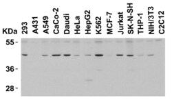
- Western Blot Validation in Human and Mouse Cell Lines. Loading: 15 µg of lysates per lane. Antibodies: PD-1 Polyclonal Antibody (Product # PA5-20350) (4 µg/mL), 1h incubation at RT in 0.05 NFDM/TBST. Secondary: Goat anti-rabbit IgG HRP conjugate at 1:10,000 dilution.

- Submitted by
- Invitrogen Antibodies (provider)
- Main image

- Experimental details
- Western Blot Validation in Rat Thymus Cell Lysate. Loading: 15 µg of lysates per lane. Antibodies: PD-1 Polyclonal Antibody (Product # PA5-20350) (1 µg/mL), 1h incubation at RT in 0.05 NFDM/TBST. Secondary: Goat anti-rabbit IgG HRP conjugate at 1:10,000 dilution.

- Submitted by
- Invitrogen Antibodies (provider)
- Main image

- Experimental details
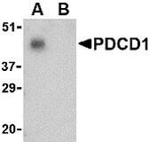
- Western Blot Validation in THP-1 Cell Lysate in the (A) absence and (B) presence of blocking peptide. Loading: 15 µg of lysates per lane. Antibodies: PD-1 Polyclonal Antibody (Product # PA5-20350) (1 µg/mL), 1h incubation at RT in 0.05 NFDM/TBST. Secondary: Goat anti-rabbit IgG HRP conjugate at 1:10,000 dilution.
Supportive validation
- Submitted by
- Invitrogen Antibodies (provider)
- Main image

- Experimental details
- Immunohistochemical analysis of paraffin-embedded human brain tissue using PD-1 Polyclonal Antibody (Product # PA5-20350) at 2.5 µg/mL. Tissue was fixed with formaldehyde and blocked with 0.1 serum for 1 h at RT; antigen retrieval was by heat mediation with a citrate buffer (pH6). Samples were incubated with primary antibody overnight at 4˚C. A goat anti-rabbit IgG H&L (HRP) at 1/250 was used as secondary. Counter stained with Hematoxylin.

- Submitted by
- Invitrogen Antibodies (provider)
- Main image

- Experimental details
- Immunohistochemical analysis of paraffin-embedded human tonsil tissue using PD-1 Polyclonal Antibody (Product # PA5-20350) at 5 µg/mL. Tissue was fixed with formaldehyde and blocked with 0.1 serum for 1 h at RT; antigen retrieval was by heat mediation with a citrate buffer (pH6). Samples were incubated with primary antibody overnight at 4˚C. A goat anti-rabbit IgG H&L (HRP) at 1/250 was used as secondary. Counter stained with Hematoxylin.

Supportive validation
- Submitted by
- Invitrogen Antibodies (provider)
- Main image

- Experimental details
- Fig. 4 Pep R in combination with anti-PD-1 reduced the expression of CXCR4-CXCL12 and PD-L1 in MC38 tumors. Representative IHC pictures for CXCR4, CXCR7, CXCL12, PD-1 and PD-L1 expression (brown staining) in MC38 collected tumors (magnification 400x), from mice treated with Pep R, anti-murine PD-1 or combined treatment showing the reduction of CXCR4, CXCL12 and PD-L1 expression in mice treated with Pep R alone and in combination with anti-PD-1

- Submitted by
- Invitrogen Antibodies (provider)
- Main image
- Experimental details
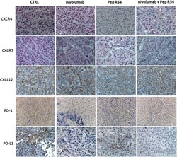
- Fig. 7 Pep R54 in combination with nivolumab reduced the expression of CXCR4-CXCL12 and PD-L1 in PES43 tumors. Representative IHC pictures (magnification 400x) for CXCR4, CXCR7 (red staining), CXCL12, PD-1 and PD-L1 expression (brown staining) in PES43 collected tumors from mice treated with Pep R54, nivolumab or combined treatment
