 Explore
 Validate
 Learn
Learn Western blot
 Immunocytochemistry
 Immunoprecipitation
Antibody data
- Antibody Data
- Antigen structure
- References [0]
- Comments [0]
- Validations
- Western blot [1]
- Immunoprecipitation [3]
Submit
Validation data
Reference
Comment
Report error
- Product number
- LS-C353523 - Provider product page

- Provider
- LSBio
- Product name
- SEMA4A / Semaphorin 4A Antibody (Internal) LS-C353523
- Antibody type
- Polyclonal
- Description
- Immunoaffinity purified
- Reactivity
- Human, Simian
- Host
- Rabbit
- Storage
- Store at -20°C. Aliquot to avoid freeze/thaw cycles.
No comments: Submit comment
Enhanced validation
- Submitted by
- LSBio (provider)
- Enhanced method
- Genetic validation
- Main image
- Experimental details
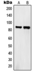
- Western blot analysis of Semaphorin 4A expression in HeLa (A); COS7 (B) whole cell lysates.

Supportive validation
- Submitted by
- LSBio (provider)
- Enhanced method
- Genetic validation
- Main image
- Experimental details
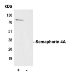
- Immunoprecipitation of Semaphorin 4A from 0.5mg Jurkat whole cell extract lysate using 5ug of Anti-Semaphorin 4A Antibody and 50ul of protein G magnetic beads (+). No antibody was added to the control (-). The antibody was incubated under agitation with Protein G beads for 10min Jurkat whole cell extract lysate diluted in RIPA buffer was added to each sample and incubated for a further 10min under agitation. Proteins were eluted by addition of 40ul SDS loading buffer and incubated for 10min at 70 C; 10ul of each sample was separated on a SDS PAGE gel transferred to a nitrocellulose membrane blocked with 5% BSA and probed with Anti-Semaphorin 4A Antibody.

- Submitted by
- LSBio (provider)
- Main image

- Experimental details
- Immunoprecipitation of Semaphorin 4A from 0.5mg Jurkat whole cell extract lysate using 5ug of Anti-Semaphorin 4A Antibody and 50ul of protein G magnetic beads (+). No antibody was added to the control (-). The antibody was incubated under agitation with Protein G beads for 10min Jurkat whole cell extract lysate diluted in RIPA buffer was added to each sample and incubated for a further 10min under agitation. Proteins were eluted by addition of 40ul SDS loading buffer and incubated for 10min at 70 C; 10ul of each sample was separated on a SDS PAGE gel transferred to a nitrocellulose membrane blocked with 5% BSA and probed with Anti-Semaphorin 4A Antibody.
