 Explore
 Validate
 Learn
Learn Western blot
Antibody data
- Antibody Data
- Antigen structure
- References [3]
- Comments [0]
- Validations
- Western blot [1]
- Immunohistochemistry [1]
Submit
Validation data
Reference
Comment
Report error
- Product number
- AF3909 - Provider product page

- Provider
- R&D Systems
- Product name
- Human/Mouse Phospho-Tie-2 (Y1102/Y1100) Antibody
- Antibody type
- Polyclonal
- Description
- Antigen Affinity-purified. Detects human and mouse Tie-2 when phosphorylated at Y1102 and Y1100, respectively.
- Reactivity
- Human, Mouse
- Host
- Rabbit
- Conjugate
- Unconjugated
- Isotype
- IgG
- Vial size
- 100 ug
- Concentration
- LYOPH
- Storage
- Use a manual defrost freezer and avoid repeated freeze-thaw cycles. 12 months from date of receipt, -20 to -70 °C as supplied. 1 month, 2 to 8 °C under sterile conditions after reconstitution. 6 months, -20 to -70 °C under sterile conditions after reconstitution.
Submitted references The Fibrin Cleavage Product Bβ(15-42) Channels Endothelial and Tubular Regeneration in the Post-acute Course During Murine Renal Ischemia Reperfusion Injury.
Flunarizine suppresses endothelial Angiopoietin-2 in a calcium - dependent fashion in sepsis.
Opposing effects of the angiopoietins on the thrombin-induced permeability of human pulmonary microvascular endothelial cells.
Fischer D, Seifen C, Baer P, Jung M, Mertens C, Scheller B, Zacharowski K, Hofmann R, Maier TJ, Urbschat A
Frontiers in pharmacology 2018;9:369
Frontiers in pharmacology 2018;9:369
Flunarizine suppresses endothelial Angiopoietin-2 in a calcium - dependent fashion in sepsis.
Retzlaff J, Thamm K, Ghosh CC, Ziegler W, Haller H, Parikh SM, David S
Scientific reports 2017 Mar 9;7:44113
Scientific reports 2017 Mar 9;7:44113
Opposing effects of the angiopoietins on the thrombin-induced permeability of human pulmonary microvascular endothelial cells.
van der Heijden M, van Nieuw Amerongen GP, van Bezu J, Paul MA, Groeneveld AB, van Hinsbergh VW
PloS one 2011;6(8):e23448
PloS one 2011;6(8):e23448
No comments: Submit comment
Supportive validation
- Submitted by
- R&D Systems (provider)
- Main image
- Experimental details
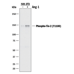
- Detection of Mouse Phospho-Tie-2 (Y1102/Y1100) by Western Blot. Western blot shows lysates of NIH-3T3 mouse embryonic fibroblast cell line transfected with mouse Tie-2 untreated (-) or treated (+) with 600 ng/mL Recombinant Human Angiopoietin-1 (Catalog # 923-AN) for 5 minutes. PVDF membrane was probed with 1 µg/mL of Rabbit Anti-Human/Mouse Phospho-Tie-2 (Y1102/Y1100) Antigen Affinity-purified Polyclonal Antibody (Catalog # AF3909) followed by HRP-conjugated Anti-Rabbit IgG Secondary Antibody (Catalog # HAF008). A specific band was detected for Phospho-Tie-2 (Y1102/1100) at approximately 150 kDa (as indicated). This experiment was conducted under reducing conditions and using Immunoblot Buffer Group 1.

Supportive validation
- Submitted by
- R&D Systems (provider)
- Main image

- Experimental details
- Tie-2 in Human Placenta. Tie-2 phosphorylated at Y1102 was detected in immersion fixed paraffin-embedded sections of human placenta using Rabbit Anti-Human/Mouse Phospho-Tie-2 (Y1102/Y1100) Antigen Affinity-purified Polyclonal Antibody (Catalog # AF3909) at 15 µg/mL overnight at 4 °C. Tissue was stained using the Anti-Rabbit HRP-DAB Cell & Tissue Staining Kit (brown; Catalog # CTS005) and counterstained with hematoxylin (blue). Lower panel shows a lack of labeling if primary antibodies are omitted and tissue is stained only with secondary antibody followed by incubation with detection reagents. View our protocol for Chromogenic IHC Staining of Paraffin-embedded Tissue Sections.
