 Explore
 Validate
 Learn
Learn Western blot
Antibody data
- Antibody Data
- Antigen structure
- References [0]
- Comments [0]
- Validations
- Western blot [1]
- Immunocytochemistry [1]
- Immunohistochemistry [1]
- Flow cytometry [1]
Submit
Validation data
Reference
Comment
Report error
- Product number
- MAB71527-100 - Provider product page

- Provider
- R&D Systems
- Product name
- Human/Mouse/Rat ATF6 Antibody
- Antibody type
- Monoclonal
- Description
- Protein A or G purified from cell culture supernatant. Detects human ATF6 in direct ELISAs. Detects human, mouse, and rat ATF6 in Western blots.
- Reactivity
- Human, Mouse, Rat
- Host
- Rabbit
- Conjugate
- Unconjugated
- Antigen sequence
P18850- Isotype
- IgG
- Antibody clone number
- 2358C
- Vial size
- 100 ug
- Storage
- Use a manual defrost freezer and avoid repeated freeze-thaw cycles. 12 months from date of receipt, -20 to -70 °C as supplied. 1 month, 2 to 8 °C under sterile conditions after reconstitution. 6 months, -20 to -70 °C under sterile conditions after reconstitution.
No comments: Submit comment
Supportive validation
- Submitted by
- R&D Systems (provider)
- Main image
- Experimental details
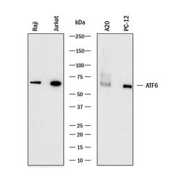
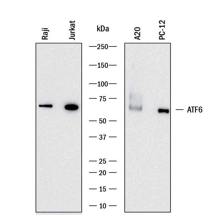
- Detection of Human, Mouse, and Rat ATF6 by Western Blot. Western blot shows lysates of Raji human Burkitt's lymphoma cell line, Jurkat human acute T cell leukemia cell line, A20 mouse B cell lymphoma cell line, and PC-12 rat adrenal pheochromocytoma cell line. PVDF membrane was probed with 0.05 µg/mL of Rabbit Anti-Human/Mouse/Rat ATF6 Monoclonal Antibody (Catalog # MAB71527) followed by HRP-conjugated Anti-Rabbit IgG Secondary Antibody (Catalog # HAF008). A specific band was detected for ATF6 at approximately 70 kDa (as indicated). This experiment was conducted under reducing conditions and using Immunoblot Buffer Group 1.
Supportive validation
- Submitted by
- R&D Systems (provider)
- Main image
- Experimental details
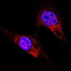
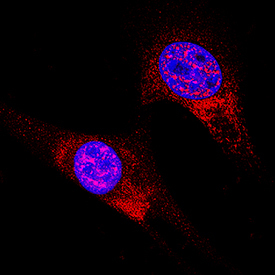
- ATF6 in A549 Human Cell Line. ATF6 was detected in immersion fixed A549 human lung carcinoma cell line using Rabbit Anti-Human/Mouse/Rat ATF6 Monoclonal Antibody (Catalog # MAB71527) at 8 µg/mL for 3 hours at room temperature. Cells were stained using the NorthernLights™ 557-conjugated Anti-Rabbit IgG Secondary Antibody (red; Catalog # NL004) and counterstained with DAPI (blue). Specific staining was localized to cytoplasm and nuclei. View our protocol for Fluorescent ICC Staining of Cells on Coverslips.
Supportive validation
- Submitted by
- R&D Systems (provider)
- Main image
- Experimental details
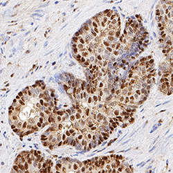
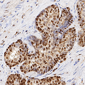
- ATF6 in Human Prostate. ATF6 was detected in immersion fixed paraffin-embedded sections of human prostate using Rabbit Anti-Human/Mouse/Rat ATF6 Monoclonal Antibody (Catalog # MAB71527) at 0.3 µg/mL for 1 hour at room temperature followed by incubation with the Anti-Rabbit IgG VisUCyte™ HRP Polymer Antibody (Catalog # VC003). Before incubation with the primary antibody, tissue was subjected to heat-induced epitope retrieval using Antigen Retrieval Reagent-Basic (Catalog # CTS013). Tissue was stained using DAB (brown) and counterstained with hematoxylin (blue). Specific staining was localized to cytoplasm and nuclei. View our protocol for IHC Staining with VisUCyte HRP Polymer Detection Reagents.
Supportive validation
- Submitted by
- R&D Systems (provider)
- Main image
- Experimental details
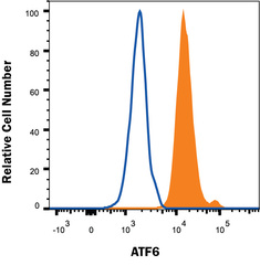
- Detection of ATF6 in Human HeLa cells by Flow Cytometry. HeLa human cervical epithelial cell line was stained with Rabbit Anti-Human/Mouse/Rat ATF6 Monoclonal Antibody (Catalog # MAB71527, filled histogram) or Rabbit IgG control antibody (Catalog # MAB1050, open histogram) followed by APC-conjugated Anti-Rabbit IgG Secondary Antibody (Catalog # F0111). To facilitate intracellular staining, cells were fixed and permeabilized with FlowX FoxP3 Fixation & Permeabilization Buffer Kit (Catalog # FC012). View our protocol for Staining Intracellular Molecules.
