Explore
 Validate
 Learn
Learn Western blot
 Immunocytochemistry
Antibody data
- Antibody Data
- Antigen structure
- References [1]
- Comments [0]
- Validations
- Immunocytochemistry [1]
- Immunohistochemistry [3]
- Chromatin Immunoprecipitation [2]
- Other assay [2]
Submit
Validation data
Reference
Comment
Report error
- Product number
- PA5-27733 - Provider product page

- Provider
- Invitrogen Antibodies
- Product name
- PTPN12 Polyclonal Antibody
- Antibody type
- Polyclonal
- Antigen
- Recombinant full-length protein
- Description
- Recommended positive controls: A549, HeLa, HepG2. Predicted reactivity: Mouse (99%), Rat (98%), Xenopus laevis (81%), Bovine (98%). Store product as a concentrated solution. Centrifuge briefly prior to opening the vial.
- Reactivity
- Human, Rat
- Host
- Rabbit
- Isotype
- IgG
- Vial size
- 100 μL
- Concentration
- 0.93 mg/mL
- Storage
- Store at 4°C short term. For long term storage, store at -20°C, avoiding freeze/thaw cycles.
Submitted references Evaluating Large Spontaneous Deletions in a Bovine Cell Line Selected for Bovine Viral Diarrhea Virus Resistance.
Workman AM, Heaton MP, Webster DA, Harhay GP, Kalbfleisch TS, Smith TPL, Falkenberg SM, Carlson DF, Sonstegard TS
Viruses 2021 Oct 25;13(11)
Viruses 2021 Oct 25;13(11)
No comments: Submit comment
Supportive validation
- Submitted by
- Invitrogen Antibodies (provider)
- Main image
- Experimental details
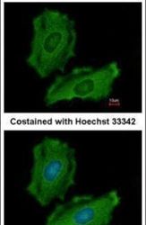
- Immunofluorescent analysis of PTPN12 in methanol-fixed HeLa cells using a PTPN12 polyclonal antibody (Product # PA5-27733) at a 1:200 dilution.

Supportive validation
- Submitted by
- Invitrogen Antibodies (provider)
- Main image
- Experimental details
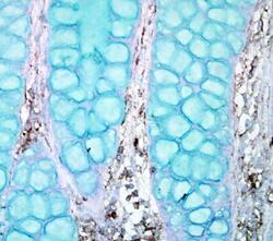
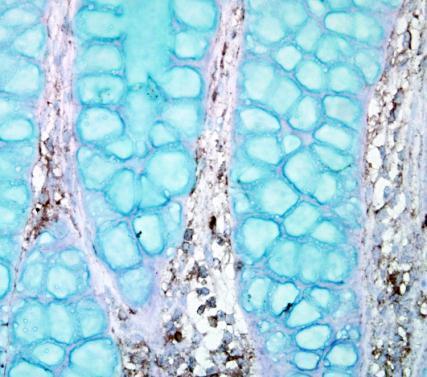
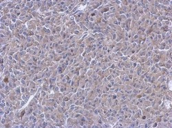
- Immunohistochemical analysis of paraffin-embedded human ulcerative colitis tissue using PTPN12 antibody (Product # PA5-27733).
- Submitted by
- Invitrogen Antibodies (provider)
- Main image
- Experimental details
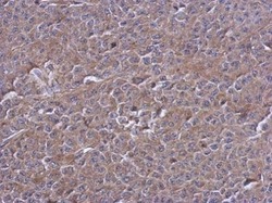
- Immunohistochemical analysis of paraffin-embedded RT2 xenograft, using PTPN12 (Product # PA5-27733) antibody at 1:500 dilution. Antigen Retrieval: EDTA based buffer, pH 8.0, 15 min.

- Submitted by
- Invitrogen Antibodies (provider)
- Main image
- Experimental details
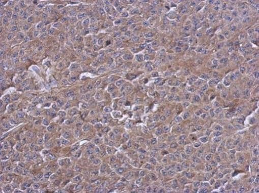
- Immunohistochemical analysis of paraffin-embedded D54 xenograft, using PTPN12 (Product # PA5-27733) antibody at 1:500 dilution. Antigen Retrieval: EDTA based buffer, pH 8.0, 15 min.
Supportive validation
- Submitted by
- Invitrogen Antibodies (provider)
- Main image
- Experimental details
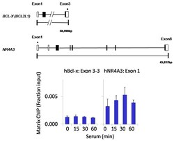
- Chromatin immunoprecipitation analysis of PTPN12 was performed using cross-linked chromatin from 1x10^6 HCT116 colon carcinoma cells treated with serum for 0, 15, 30, and 60 minutes. Immunoprecipitation was performed using a multiplex microplate Matrix ChIP assay (see reference for Matrix ChIP protocol: http://www.ncbi.nlm.nih.gov/pubmed/22098709) with 1.0 µL/100 µL well volume of a PTPN12 polyclonal antibody (Product # PA5-27733). Chromatin aliquots from ~1 x 10⁵ cells were used per ChIP pull-down. Quantitative PCR data were done in quadruplicate using 1 µL of eluted DNA in 2 µL SYBR real-time PCR reactions containing primers to amplify exon 3-3 of human Bcl-x (hBcl-x) or exon 1 of human NR4A3 (hNR4A3). PCR calibration curves were generated for each primer pair from a dilution series of sheared total genomic DNA. Quantitation of immunoprecipitated chromatin is presented as signal relative to the total amount of input chromatin. Results represent the mean +/- SEM for three experiments. Schematic representations of the Bcl-x and NR4A3 loci are shown above the data where boxes represent exons (black boxes = translated regions, white boxes = untranslated regions). Regions amplified by Bcl-x and NR4A3 primers are represented by black bars. Data courtesy of the Innovators Program.

- Submitted by
- Invitrogen Antibodies (provider)
- Main image

- Experimental details
- Chromatin immunoprecipitation analysis of PTPN12 was performed using cross-linked chromatin from 1x10^6 HCT116 colon carcinoma cells treated with serum for 0, 15, 30, and 60 minutes. Immunoprecipitation was performed using a multiplex microplate Matrix ChIP assay (see reference for Matrix ChIP protocol: http://www.ncbi.nlm.nih.gov/pubmed/22098709) with 1.0 µL/100 µL well volume of a PTPN12 polyclonal antibody (Product # PA5-27733). Chromatin aliquots from ~1 x 10⁵ cells were used per ChIP pull-down. Quantitative PCR data were done in quadruplicate using 1 µL of eluted DNA in 2 µL SYBR real-time PCR reactions containing primers to amplify exon 3-3 of human Bcl-x (hBcl-x) or exon 1 of human NR4A3 (hNR4A3). PCR calibration curves were generated for each primer pair from a dilution series of sheared total genomic DNA. Quantitation of immunoprecipitated chromatin is presented as signal relative to the total amount of input chromatin. Results represent the mean +/- SEM for three experiments. Schematic representations of the Bcl-x and NR4A3 loci are shown above the data where boxes represent exons (black boxes = translated regions, white boxes = untranslated regions). Regions amplified by Bcl-x and NR4A3 primers are represented by black bars. Data courtesy of the Innovators Program.

Supportive validation
- Submitted by
- Invitrogen Antibodies (provider)
- Main image
- Experimental details
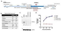
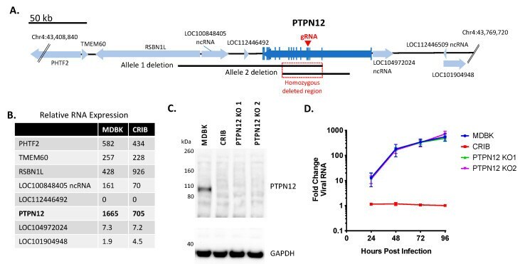
- Figure 2 Knockout of PTPN12 does not significantly impact BVDV infection in MDBK cells. Panel ( A ), map of bovine PTPN12 and surrounding genes: blue arrows, genes (open reading frames); vertical blue lines, coding regions (exons); black lines, heterozygous deletions in two alleles of PTPN12 on chromosome 4; red square, homozygous deleted region (33,808 bp); red arrow, exon target of PTPN12 gRNA. Panel ( B ), RNASeq analyses of relative RNA transcript abundance in MDBK and CRIB cells (trimmed mean of M (TMM) normalization value). Panel ( C ), western blot of SDS-PAGE for PTPN12 and GAPDH (loading control). Panel ( D ), Multistep virus growth curve. Cells were infected with BVDV strain NADL (MOI 0.01). Cells were collected and processed 0-96 h post-infection for quantitation of viral RNA using RT-qPCR. Results represent the mean +- standard deviation ( n = 3).
- Submitted by
- Invitrogen Antibodies (provider)
- Main image
- Experimental details
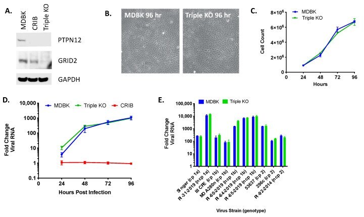
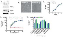
- Figure 5 Triple KO of PTPN12 , GRID2 , and RABGAP1L genes does not impact BVDV infection in MDBK cells. Panel ( A ) western blot of SDS-PAGE for PTPN12, GRID2, and GAPDH (loading control). Panel ( B ) MDBK and MDBK-TKO cell morphology (10x). Panel ( C ) Cell growth in T-25 flasks. Panel ( D ) Multistep virus growth curve. Cells were infected with BVDV strain NADL (MOI 0.01). Cells were collected and processed 0-96 h post-infection for quantitation of viral RNA using RT-qPCR. Results represent the mean +- standard deviation ( n = 3). Panel ( E ) Cells were infected with various cytopathic (cp) and non-cytopathic (ncp) BVDV isolates (MOI = 0.01) and collected at 0 and 96 h post-infection for quantitation of viral RNA using RT-qPCR. Results represent means at 96 h +- standard deviation ( n = 3).