Explore
 Validate
 Learn
Learn Western blot
Antibody data
- Antibody Data
- Antigen structure
- References [0]
- Comments [0]
- Validations
- Western blot [1]
- Immunocytochemistry [1]
- Flow cytometry [1]
Submit
Validation data
Reference
Comment
Report error
- Product number
- ABIN2508732 - Provider product page

- Provider
- antibodies-online
- Product name
- anti-Chemokine (C Motif) Ligand 1 (XCL1) antibody (Biotin)
- Antibody type
- Polyclonal
- Antigen
- Other
- Description
- Produced from sera of rabbits pre-immunized with highly pure (>98%) recombinant hLymphotactin. Anti-Human Lymphotactin specific antibody was purified by affinity chromatography and then biotinylated.
- Reactivity
- Human
- Host
- Rabbit
- Conjugate
- Biotin
- Vial size
- 50 μg
- Storage
- -20°C
No comments: Submit comment
Supportive validation
- Submitted by
- antibodies-online (provider)
- Main image
- Experimental details
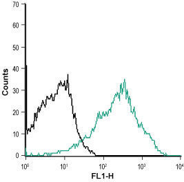
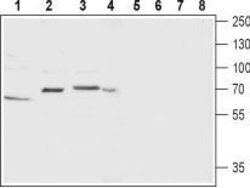
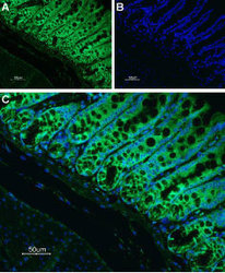
- Western blot analysis of rat ovary (lanes 1 and 5), rat brain (lanes 2 and 6), mouse brain membrane (lanes 3 and 7) and SH-SY5Y (lanes 4 and 8) lysate: 1-4. Anti-Neurokinin Receptor 1 (NK1) (extracellular) antibody (ABIN2511130), (1:200). 5-8. Anti-Neurokinin Receptor 1 (NK1) (extracellular) antibody, preincubated with the control peptide antigen. Expression of Neurokinin Receptor 1 in rat colon Immunohistochemical staining of rat colon paraffin-embedded section using Anti-Neurokinin Receptor 1 (NK1) (extracellular) antibody (ABIN2511130), (1:100) followed by goat anti-rabbit-AlexaFluor-488 secondary antibody. A. NK1 labeling appears in the tubular glands of the mucosa layer. Note that the smooth muscle and lamina propria do not stain. B. Nuclear staining using DAPI as the counterstain. C. Merged images of A and B. Indirect flow cytometry analysis of MEG-O1 living cells: ? Unstained cells. ? Cells + Anti-Anti-Neurokinin Receptor 1 (NK1) (extracellular) antibody (ABIN2511130), (5-10 μg/1x106 cells).

Supportive validation
- Submitted by
- antibodies-online (provider)
- Main image
- Experimental details
- Image(s): Immunofluorescence

Supportive validation
- Submitted by
- antibodies-online (provider)
- Main image

- Experimental details
- Image(s): Flow cytometry