 Explore
 Validate
 Learn
Learn Western blot
 Immunocytochemistry
Antibody data
- Antibody Data
- Antigen structure
- References [0]
- Comments [0]
- Validations
- Western blot [8]
- Immunocytochemistry [1]
- Immunoprecipitation [1]
- Immunohistochemistry [2]
Submit
Validation data
Reference
Comment
Report error
- Product number
- GTX102619 - Provider product page

- Provider
- GeneTex
- Product name
- ROCK2 antibody
- Antibody type
- Polyclonal
- Reactivity
- Human, Mouse, Rat
- Host
- Rabbit
No comments: Submit comment
Enhanced validation
Supportive validation
- Submitted by
- GeneTex (provider)
- Enhanced method
- Genetic validation
- Main image

- Experimental details
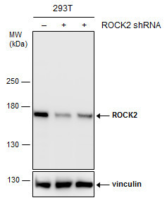
- Non-transfected (¡V) and transfected (+) 293T whole cell extracts (30 ?g) were separated by 5% SDS-PAGE, and the membrane was blotted with ROCK2 antibody (GTX102619) diluted at 1:2000.
Supportive validation
- Submitted by
- GeneTex (provider)
- Main image

- Experimental details
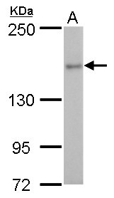
- ROCK2 antibody detects ROCK2 protein by western blot analysis.A. 30 £gg Rat2 whole cell lysate/extract5 % SDS-PAGEROCK2 antibody (GTX102619) dilution: 1:1000
- Submitted by
- GeneTex (provider)
- Main image

- Experimental details
- ROCK2 antibody detects ROCK2 protein by western blot analysis.A. 30 £gg Neuro2A whole cell lysate/extract B. 30 £gg GL261 whole cell lysate/extractC. 30 £gg C8D30 whole cell lysate/extractD. 30 £gg NIH-3T3 whole cell lysate/extractE. 30 £gg BCL-1 whole cell lysate/extract F. 30 £gg Raw 264.7 whole cell lysate/extract G. 30 £gg C2Cl2 whole cell lysate/extract5 % SDS-PAGEROCK2 antibody (GTX102619) dilution: 1:1000

- Submitted by
- GeneTex (provider)
- Main image

- Experimental details
- ROCK2 antibody detects ROCK2 protein by western blot analysis.A. 30 £gg A549 whole cell lysate/extract B. 30 £gg H1299 whole cell lysate/extract C. 30 £gg HCT116 whole cell lysate/extract5 % SDS-PAGEROCK2 antibody (GTX102619) dilution: 1:1000

- Submitted by
- GeneTex (provider)
- Main image

- Experimental details
- ROCK2 antibody detects ROCK2 protein by western blot analysis. Various whole cell extracts (30 £gg) were separated by 5% SDS-PAGE, and the membrane was blotted with ROCK2 antibody (GTX102619) diluted at a dilution of 1:1000.

- Submitted by
- GeneTex (provider)
- Main image

- Experimental details
- ROCK2 antibody detects ROCK2 protein by western blot analysis. Various whole cell extracts (30 ?g) were separated by 5% SDS-PAGE, and the membrane was blotted with ROCK2 antibody (GTX102619) diluted by 1:1000.
- Validation comment
- WB

- Submitted by
- GeneTex (provider)
- Main image

- Experimental details
- Various whole cell extracts (30 ?g) were separated by 5% SDS-PAGE, and the membranes were blotted with ROCK2 antibody (GTX102619) diluted at 1:1000 and competitor's antibody (sc-1851) diluted at 1:100. The HRP-conjugated anti-rabbit IgG antibody (GTX213110-01) was used to detect the primary antibody.*The competitor is not affiliated with GeneTex and does not endorse this product.

- Submitted by
- GeneTex (provider)
- Main image
- Experimental details
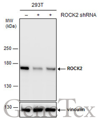
- Non-transfected (¡V) and transfected (+) 293T whole cell extracts (30 ?g) were separated by 5% SDS-PAGE, and the membrane was blotted with ROCK2 antibody (GTX102619) diluted at 1:2000.

Supportive validation
- Submitted by
- GeneTex (provider)
- Main image

- Experimental details
- ROCK2 antibody detects ROCK2 protein at cytoplasm by immunofluorescent analysis.Sample: A431 cells were fixed in ice-cold MeOH for 5 min.Green: ROCK2 protein stained by ROCK2 antibody (GTX102619) diluted at 1:500.Blue: Hoechst 33342 staining.

Supportive validation
- Submitted by
- GeneTex (provider)
- Main image

- Experimental details
- Immunoprecipitation of ROCK2 protein from HeLa whole cell extracts using 5 £gg of ROCK2 antibody (GTX102619).Western blot analysis was performed using ROCK2 antibody (GTX102619).EasyBlot anti-Rabbit IgG (GTX221666-01) was used as a secondary reagent.

Supportive validation
- Submitted by
- GeneTex (provider)
- Main image

- Experimental details
- ROCK2 antibody detects ROCK2 protein at cytosol on mouse kidney by immunohistochemical analysis. Sample: Paraffin-embedded mouse kidney. ROCK2 antibody (GTX102619) dilution: 1:500.

- Submitted by
- GeneTex (provider)
- Main image
- Experimental details
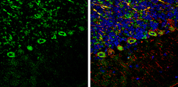
- ROCK2 antibody detects ROCK2 protein expression by immunohistochemical analysis.Sample: Frozen-sectioned adult mouse cerebellum. Green: ROCK2 protein stained by ROCK2 antibody (GTX102619) diluted at 1:250.Red: NF-H, stained by NF-H antibody [GT114] (GTX634289) diluted at 1:500.Blue: Fluoroshield with DAPI (GTX30920).
