Explore
 Validate
 Learn
Learn Western blot
Antibody data
- Antibody Data
- Antigen structure
- References [0]
- Comments [0]
- Validations
- Western blot [2]
- Immunocytochemistry [1]
- Immunohistochemistry [1]
- Flow cytometry [1]
Submit
Validation data
Reference
Comment
Report error
- Product number
- MAB7680-100 - Provider product page

- Provider
- R&D Systems
- Product name
- Human/Rat EGLN1/PHD2 Antibody
- Antibody type
- Monoclonal
- Description
- Protein A or G purified from cell culture supernatant. Detects human EGLN/PHD2 in direct ELISAs. Detects human and rat EGLN/PHD2 in Western blots. In direct ELISAs, no cross-reactivity with human PHD1 and PHD3 is observed.
- Reactivity
- Human, Rat
- Host
- Rabbit
- Conjugate
- Unconjugated
- Antigen sequence
Q9GZT9- Isotype
- IgG
- Antibody clone number
- 2445B
- Vial size
- 100 ug
- Storage
- Use a manual defrost freezer and avoid repeated freeze-thaw cycles. 12 months from date of receipt, -20 to -70 °C as supplied. 1 month, 2 to 8 °C under sterile conditions after reconstitution. 6 months, -20 to -70 °C under sterile conditions after reconstitution.
No comments: Submit comment
Supportive validation
- Submitted by
- R&D Systems (provider)
- Main image

- Experimental details
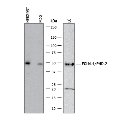
- Detection of Human and Rat EGLN1/PHD2 by Western Blot. Western blot shows lysates of HEK293T human embryonic kidney cell line, PC-3 human prostate cancer cell line, and L6 rat myoblast cell line. PVDF membrane was probed with 1 µg/mL of Rabbit Anti-Human/Rat EGLN1/PHD2 Monoclonal Antibody (Catalog # MAB7680) followed by HRP-conjugated Anti-Rabbit IgG Secondary Antibody (Catalog # HAF008). A specific band was detected for EGLN1/PHD2 at approximately 49 kDa (as indicated). This experiment was conducted under reducing conditions and using Immunoblot Buffer Group 1.
- Submitted by
- R&D Systems (provider)
- Main image
- Experimental details
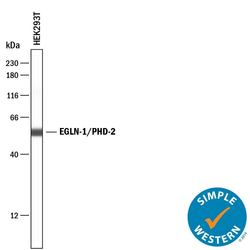
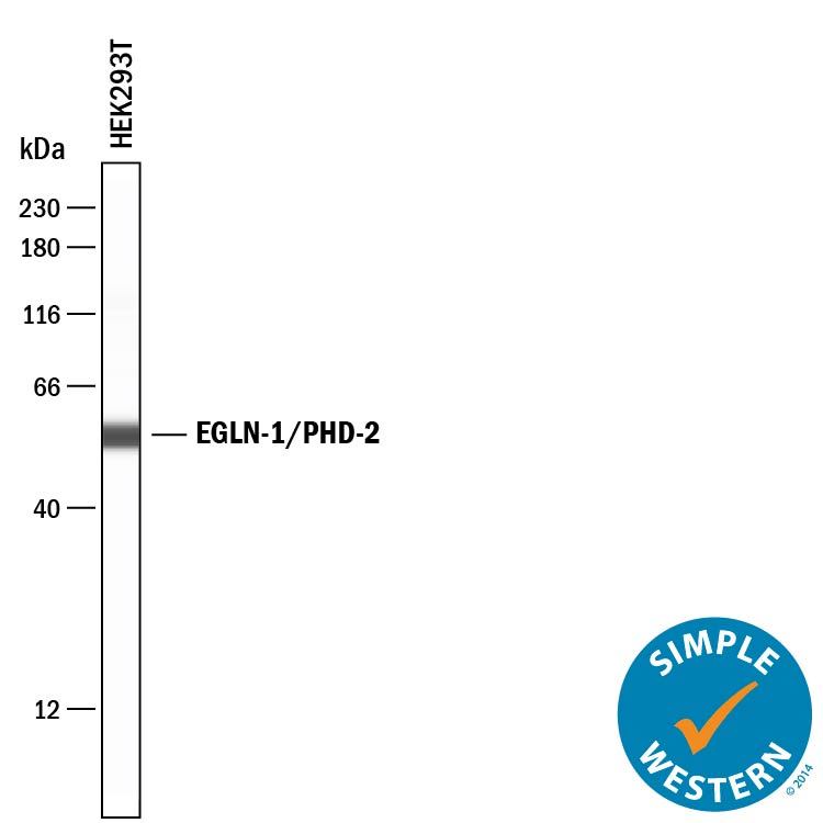
- Detection of Human EGLN1/PHD2 by Simple WesternTM. Simple Western lane view shows lysates of HEK293T human embryonic kidney cell line, loaded at 0.2 mg/mL. A specific band was detected for EGLN1/PHD2 at approximately 55 kDa (as indicated) using 10 µg/mL of Rabbit Anti-Human/Rat EGLN1/PHD2 Monoclonal Antibody (Catalog # MAB7680). This experiment was conducted under reducing conditions and using the 12-230 kDa separation system.
Supportive validation
- Submitted by
- R&D Systems (provider)
- Main image

- Experimental details
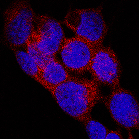
- EGLN1/PHD2 in WM-115 Human Cell Lines. EGLN1/PHD2 was detected in immersion fixed WM-115 human malignant melanoma cell line using Rabbit Anti-Human/Rat EGLN1/PHD2 Monoclonal Antibody (Catalog # MAB7680) at 3 µg/mL for 3 hours at room temperature. Cells were stained using the NorthernLights™ 557-conjugated Anti-Rabbit IgG Secondary Antibody (red; Catalog # NL004) and counterstained with DAPI (blue). Specific staining was localized to cell cytoplasm and nuclei. View our protocol for Fluorescent ICC Staining of Cells on Coverslips.
Supportive validation
- Submitted by
- R&D Systems (provider)
- Main image
- Experimental details
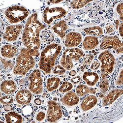
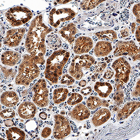
- EGLN1/PHD2 in Human Kidney. EGLN1/PHD2 was detected in immersion fixed paraffin-embedded sections of human kidney using Rabbit Anti-Human/Rat EGLN1/PHD2 Monoclonal Antibody (Catalog # MAB7680) at 3 µg/mL for 1 hour at room temperature followed by incubation with the Anti-Rabbit IgG VisUCyte™ HRP Polymer Antibody (Catalog # VC003). Before incubation with the primary antibody, tissue was subjected to heat-induced epitope retrieval using Antigen Retrieval Reagent-Basic (Catalog # CTS013). Tissue was stained using DAB (brown) and counterstained with hematoxylin (blue). Specific staining was localized to cell cytoplasm and nuclei. View our protocol for IHC Staining with VisUCyte HRP Polymer Detection Reagents.
Supportive validation
- Submitted by
- R&D Systems (provider)
- Main image

- Experimental details
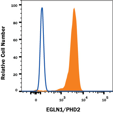
- Detection of EGLN1/PHD2 in Human Jurkat cell line by Flow Cytometry. Human Jurkat T Cell Leukemia Cell Line was stained with Rabbit Anti-Human/Rat EGLN1/PHD2 Monoclonal Antibody (Catalog # MAB7680, filled histogram) or Rabbit IgG Isotype Control Antibody (Catalog # MAB1050, open histogram) followed by Phycoerythrin-conjugated Anti-Rabbit IgG Secondary Antibody (Catalog # F0110). To facilitate intracellular staining, cells were fixed and permeabilized with FlowX FoxP3 Fixation & Permeabilization Buffer Kit (Catalog # FC012). View our protocol for Staining Membrane-associated Proteins.