 Explore
 Validate
 Learn
Learn Western blot
 Immunoprecipitation
Antibody data
- Antibody Data
- Antigen structure
- References [1]
- Comments [0]
- Validations
- Western blot [5]
- Other assay [4]
Submit
Validation data
Reference
Comment
Report error
- Product number
- PA5-34733 - Provider product page

- Provider
- Invitrogen Antibodies
- Product name
- MLKL Polyclonal Antibody
- Antibody type
- Polyclonal
- Antigen
- Recombinant protein fragment
- Description
- Recommended positive controls: THP-1, Jurkat, HCT116, PC-3. IHC notes, Requires antigen retrieval using heat mediated 10mM Citrate buffer (pH6.0) or Tris-EDTA buffer (pH8.0) Store product as a concentrated solution. Centrifuge briefly prior to opening the vial.
- Reactivity
- Human
- Host
- Rabbit
- Isotype
- IgG
- Vial size
- 100 µL
- Concentration
- 0.52 mg/mL
- Storage
- Store at 4°C short term. For long term storage, store at -20°C, avoiding freeze/thaw cycles.
Submitted references Downregulation of RIP3 Improves the Protective Effect of ATF6 in an Acute Liver Injury Model.
Huang MY, Wan DW, Deng J, Guo WJ, Huang Y, Chen H, Xu DL, Jiang ZG, Xue Y, He YH
BioMed research international 2021;2021:8717565
BioMed research international 2021;2021:8717565
No comments: Submit comment
Supportive validation
- Submitted by
- Invitrogen Antibodies (provider)
- Main image
- Experimental details
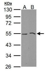
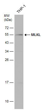
- Western blot analysis of MLKL using 30 µg of A) THP-1 and B) NCI-H929 lysate. Samples were loaded onto a 10% SDS-PAGE gel and probed with a MLKL polyclonal antibody (Product # PA5-34733) at a dilution of 1:1000.
- Submitted by
- Invitrogen Antibodies (provider)
- Main image
- Experimental details
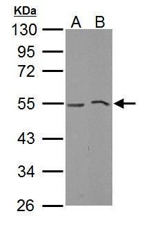
- Western Blot analysis of MLKL was performed by separating 30 µg of various whole cell extracts by 10% SDS-PAGE. Proteins were transferred to a membrane and probed with a MLKL Polyclonal Antibody (Product # PA5-34733) at a dilution of 1:500 and a HRP-conjugated anti-rabbit IgG secondary antibody.
- Submitted by
- Invitrogen Antibodies (provider)
- Main image

- Experimental details
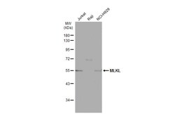
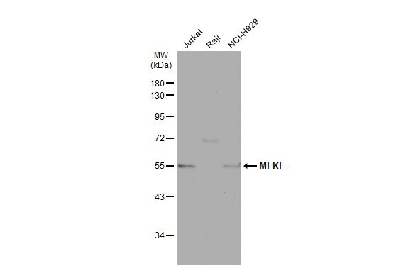
- Western blot analysis of MLKL was performed by separating 30 µg of whole cell extract by 12% SDS-PAGE. Proteins were transferred to a membrane and probed with a MLKL Polyclonal Antibody (Product # PA5-34733) at a dilution of 1:500. The HRP-conjugated anti-rabbit IgG antibody was used to detect the primary antibody.
- Submitted by
- Invitrogen Antibodies (provider)
- Main image
- Experimental details
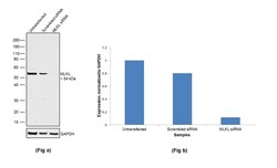
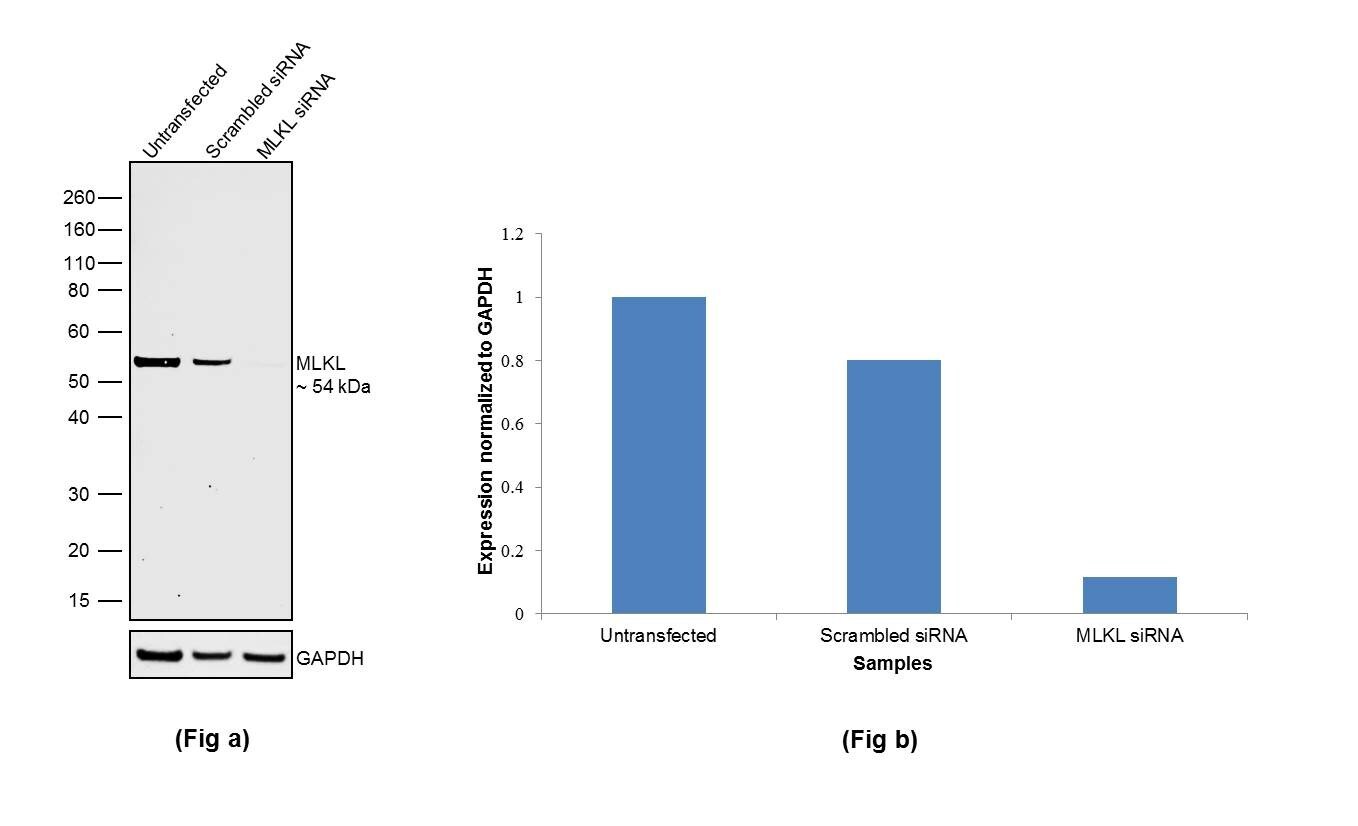
- Knockdown of MLKL was achieved by transfecting HeLa with MLKL specific siRNAs (Silencer® select Product # s200360, s47087 ). Western blot analysis (Fig. a) was performed using Membrane enriched cell extracts from the MLKL knockdown cells (lane 3), non-specific scrambled siRNA transfected cells (lane 2) and untransfected cells (lane 1). The blot was probed with MLKL Polyclonal Antibody (Product # PA5-34733, 1:2000 dilution) and Goat anti-Rabbit IgG (H+L) Superclonal™ Secondary Antibody, HRP (Product # A27036, 0.25µg/ml, 1:4000 dilution). Densitometric analysis of this western blot is shown in histogram (Fig. b). Decrease in signal upon siRNA mediated knock down confirms that antibody is specific to MLKL.
- Submitted by
- Invitrogen Antibodies (provider)
- Main image

- Experimental details
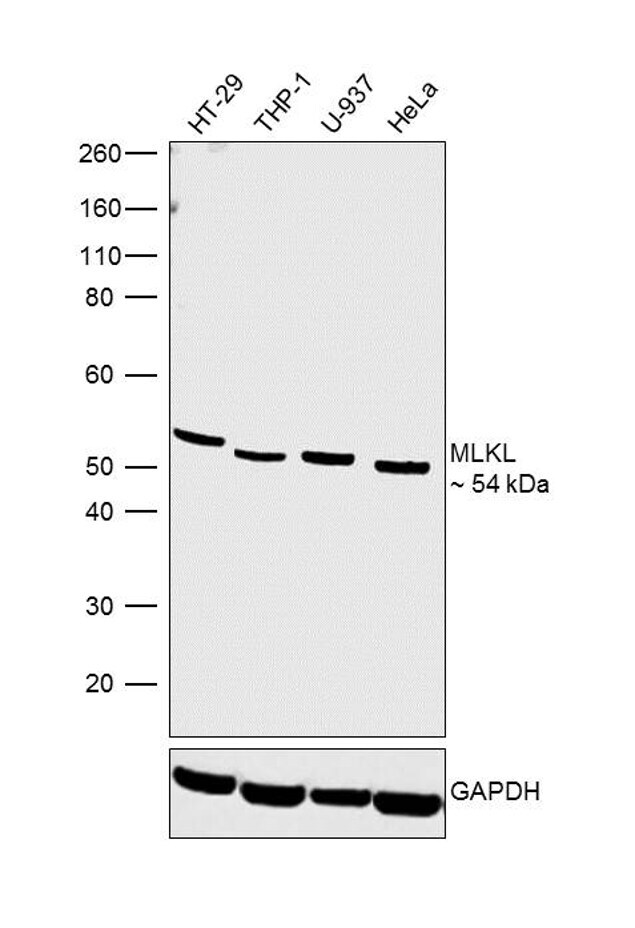
- Western blot was performed using MLKL Polyclonal Antibody (Product # PA5-34733) and a 54kDa band corresponding to MLKL was observed across cell lines tested. Membrane enriched cell extracts (30 µg lysate) of HT-29 (Lane 1), THP-1 (Lane 2), U-937 (Lane 3) and HeLa (Lane 4) were electrophoresed using NuPAGE® 4-12 % Bis-Tris gel (Product # NP0322BOX). Resolved proteins were then transferred onto a nitrocellulose membrane (Product # IB23001) by iBlot® 2 Dry Blotting System (Product # IB21001).The blot was probed with the primary antibody (1:2000 dilution) and detected by chemiluminescence with Goat Anti-Rabbit IgG Secondary Antibody, HRP conjugate (Product # A27036, 1:4000 dilution) using the iBright FL 1000 (Product # A32752). Chemiluminescent detection was performed using Novex® ECL Chemiluminescent Substrate Reagent Kit (Product # WP20005).
Supportive validation
- Submitted by
- Invitrogen Antibodies (provider)
- Main image
- Experimental details
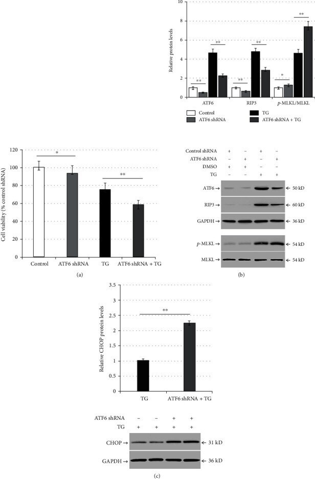
- Figure 2 ATF6 silencing aggravates TG-induced necroptosis and ER stress and reduces RIP3 expression in LO2 cells. LO2 cells were infected with control shRNA or ATF6 shRNA for 48 h, and then they were incubated with DMSO or TG (0.5 mu mol/L) for another 24 h: (a) comparison of cell viability between the control group (control shRNA + DMSO), the ATF6 shRNA group (ATF6 shRNA + DMSO), the TG group (control shRNA + TG), and the ATF6 shRNA + TG group in LO2 cells; (b) bar chart representing the ATF6, RIP3, and p-MLKL protein expression and representative western blotting analyzing the protein expression among the different experimental groups; (c) bar chart representing CHOP protein expression and representative western blotting evaluating the protein expression among the TG group and the ATF6 shRNA + TG group. * p < 0.05 and ** p < 0.01 versus the control group or the TG group.
- Submitted by
- Invitrogen Antibodies (provider)
- Main image
- Experimental details
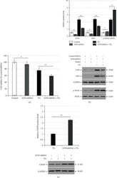
- Figure 3 RIP3 silencing alleviates TG-induced necroptosis and ER stress in LO2 cells. LO2 cells were infected with control shRNA or RIP3 shRNA for 48 h, and then they were incubated with DMSO or TG (0.5 mu mol/L) for another 24 h: (a) comparison of cell viability between the control group (control shRNA + DMSO), the RIP3 shRNA group (RIP3 shRNA + DMSO), the TG group (control shRNA + TG), and the RIP3 shRNA + TG group in LO2 cells; (b) bar chart representing the RIP3 and p-MLKL protein expression and representative western blotting analyzing the protein expression among the different experimental groups; (c) bar chart representing CHOP protein expression and representative western blotting demonstrating the protein expression among the TG group and the ATF6 shRNA + TG group. ** p < 0.01 versus the control group or the TG group.

- Submitted by
- Invitrogen Antibodies (provider)
- Main image
- Experimental details
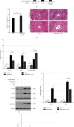
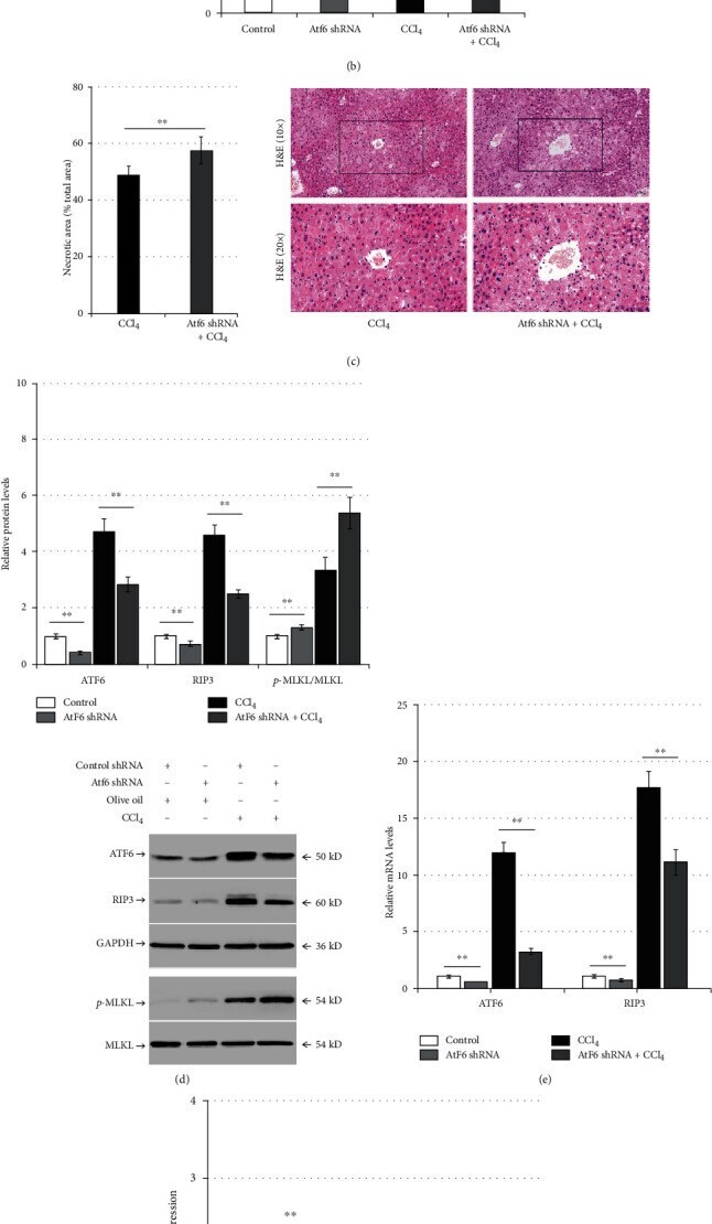
- Figure 5 Atf6 knockdown aggravates liver injury and ER stress and reduces RIP3 expression in CCl 4 -induced mice. Mice were pretreated with control shRNA or Atf6 shRNA for 6 weeks, then they were injected with olive oil or CCl 4 for 24 h ( n = 12): (a) the enzymatic rate method demonstrating the changes of serum ALT levels in the control group (control shRNA + olive oil), the Atf6 shRNA group (Atf6 shRNA + olive oil), the CCl 4 group (control shRNA + CCl 4 ), and the Atf6 shRNA + CCl 4 group; (b) serum TBil levels measured using the diazonium method in the different experimental groups; (c) H&E staining representing pathological changes in liver tissue and bar charts representing the proportion of necrotic liver tissue area; (d) western blot analysis of intrahepatic ATF6, RIP3, and p-MLKL expression among the different experimental groups; (e) qPCR analysis demonstrating the relative ATF6 and RIP3 expression among the different experimental groups; (f) western blot analysis of intrahepatic caspase-12 and CHOP expression level in the CCl 4 group and the Atf6 shRNA + CCl 4 group. ** p < 0.01 versus the control shRNA group or the control shRNA + CCl 4 group.
- Submitted by
- Invitrogen Antibodies (provider)
- Main image
- Experimental details
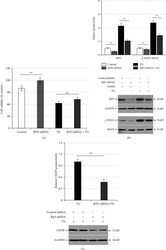
- Figure 6 RIP3 downregulation mitigates liver injury and ER stress in CCl 4 -induced mice. Mice were administered the control shRNA or Rip3 shRNA, then they were injected with olive oil or CCl 4 for another 24 h ( n = 12): (a) the enzymatic rate method to detect the changes of serum ALT levels in the control group (control shRNA + olive oil), the Rip3 shRNA group (Rip3 shRNA + olive oil), the CCl 4 group (control shRNA + CCl 4 ), and the Rip3 shRNA + CCl 4 group; (b) serum TBil levels were measured using the diazonium method among the different experimental groups; (c) H&E staining representing pathological changes in liver tissue and bar charts representing the proportion of necrotic liver tissue area; (d) western blotting examining the intrahepatic RIP3, and p-MLKL expression among the different experimental groups; (e) western blot analysis of intrahepatic caspase-12 and CHOP expression in the CCl 4 group and the Rip3 shRNA + CCl 4 group. ** p < 0.01 versus the control group or the control shRNA + CCl 4 group.
