Explore
 Validate
 Learn
Learn Western blot
Antibody data
- Antibody Data
- Antigen structure
- References [4]
- Comments [0]
- Validations
- Western blot [2]
- Other assay [4]
Submit
Validation data
Reference
Comment
Report error
- Product number
- PA5-43960 - Provider product page

- Provider
- Invitrogen Antibodies
- Product name
- MLKL Polyclonal Antibody
- Antibody type
- Polyclonal
- Antigen
- Synthetic peptide
- Description
- Peptide sequence: DVWKELSLLL QVEQRMPVSP ISQGASWAQE DQQDADEDRR AFQMLRRDNE
- Concentration
- 0.5 mg/mL
Submitted references Cell death inhibitors protect against brain damage caused by cardiac ischemia/reperfusion injury.
Serum amyloid A3 is required for caerulein-induced acute pancreatitis through induction of RIP3-dependent necroptosis.
Pomelo peel oil suppresses TNF-α-induced necroptosis and cerebral ischaemia-reperfusion injury in a rat model of cardiac arrest.
Would Colloidal Gold Nanocarriers Present An Effective Diagnosis Or Treatment For Ischemic Stroke?
Liao S, Apaijai N, Luo Y, Wu J, Chunchai T, Singhanat K, Arunsak B, Benjanuwattra J, Chattipakorn N, Chattipakorn SC
Cell death discovery 2021 Oct 23;7(1):312
Cell death discovery 2021 Oct 23;7(1):312
Serum amyloid A3 is required for caerulein-induced acute pancreatitis through induction of RIP3-dependent necroptosis.
Yang X, Li R, Xu L, Qian F, Sun L
Immunology and cell biology 2021 Jan;99(1):34-48
Immunology and cell biology 2021 Jan;99(1):34-48
Pomelo peel oil suppresses TNF-α-induced necroptosis and cerebral ischaemia-reperfusion injury in a rat model of cardiac arrest.
Wang W, Xie L, Zou X, Hu W, Tian X, Zhao G, Chen M
Pharmaceutical biology 2021 Dec;59(1):401-409
Pharmaceutical biology 2021 Dec;59(1):401-409
Would Colloidal Gold Nanocarriers Present An Effective Diagnosis Or Treatment For Ischemic Stroke?
Amani H, Mostafavi E, Alebouyeh MR, Arzaghi H, Akbarzadeh A, Pazoki-Toroudi H, Webster TJ
International journal of nanomedicine 2019;14:8013-8031
International journal of nanomedicine 2019;14:8013-8031
No comments: Submit comment
Supportive validation
- Submitted by
- Invitrogen Antibodies (provider)
- Main image
- Experimental details
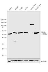
- Western blot analysis was performed on membrane enriched cell extracts (30 µg lysate) of HeLa (Lane 1), SW480 (Lane 2), U-937 (Lane 3), HT-29 (Lane 4), Mouse Brain (Lane 5) and Mouse Liver (Lane 6). The blot was probed with Anti-MLKL Polyclonal Antibody (Product # PA5-43960, 1µg/ml dilution) and detected by chemiluminescence using Goat anti-Rabbit IgG (H+L) Superclonal™ Secondary Antibody, HRP conjugate (Product # A27036, 0.25 µg/ml, 1:4000 dilution). A 55 kDa band corresponding to MLKL was observed across all the cell lines and tissues tested except Mouse Brain. A non-specific band at ~150 kDa was observed in Mouse Brain tissue lysate.

- Submitted by
- Invitrogen Antibodies (provider)
- Main image
- Experimental details
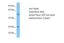
- Western blot analysis of human 293T whole cell lysate using an anti-MLKL polyclonal antibody (Product # PA5-43960).

Supportive validation
- Submitted by
- Invitrogen Antibodies (provider)
- Main image
- Experimental details
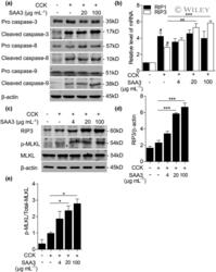
- 6 Figure rSAA3 induces RIP3 expression and MLKL phosphorylation. (a) The protein levels of pro- and cleaved caspase-3, caspase-8 and caspase-9 in AR42J cells under CCK stimulation with or without rSAA3 were measured by western blot. (b) The mRNA levels of RIP1 and RIP3 in AR42J cells under CCK stimulation with or without rSAA3 were measured by quantitative RT-PCR. The results are shown as the relative levels of gene transcripts, with those of the no treatment group set as 1. (c) The protein levels of RIP3 and p-MLKL in AR42J cells under CCK stimulation with or without rSAA3 were measured by western blot. beta-actin and total MLKL were used as loading control. (d , e) The quantification of the blots in c is shown. All quantitative data shown are means +- s.e.m. based on triplicate measurements. # P < 0.05 versus the control group, * P < 0.05, **P < 0.01, *** P < 0.001 versus the CCK group. CCK, cholecystokinin; MLKL, mixed lineage kinase domain-like protein; mRNA, messenger RNA; RIP1, RIP3, receptor-interacting protein 1; RIP3, receptor-interacting protein 3; rSAA3, recombinant serum amyloid A 3; RT-PCR, real-time PCR.

- Submitted by
- Invitrogen Antibodies (provider)
- Main image
- Experimental details
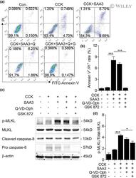
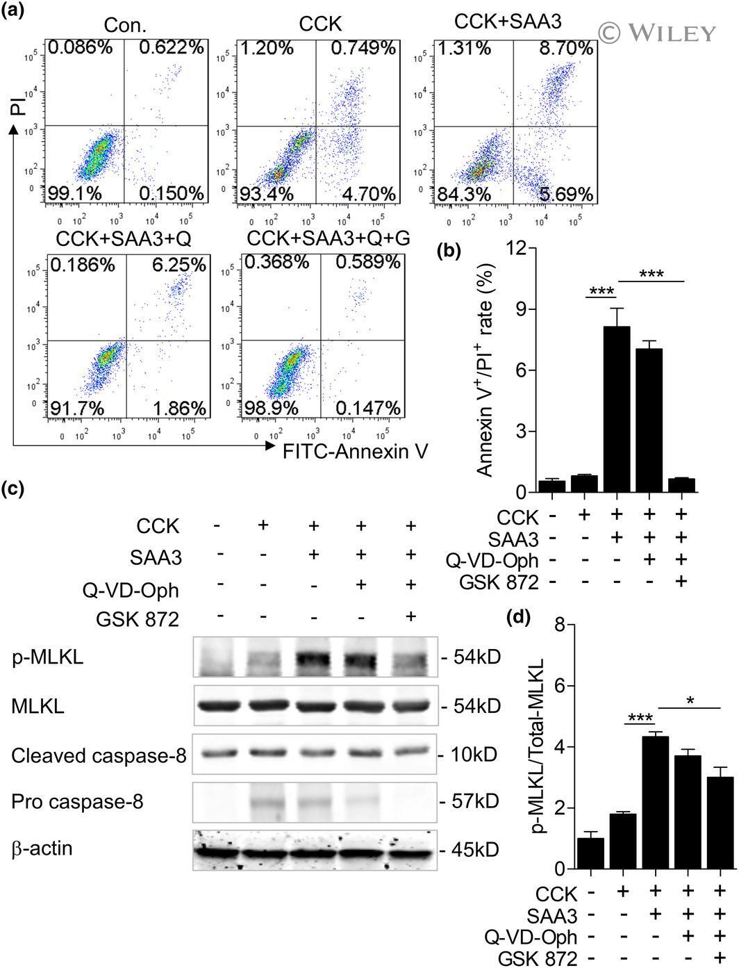
- 8 Figure rSAA3 induced acinar cell necroptosis through an RIP3-dependent pathway. (a) Caspase inhibitor (Q-VD-Oph) was added to AR42J cells 1 h prior to CCK treatment with or without rSAA3 stimulation. RIP3 inhibitor was added to AR42J cells 30 min prior to treatment with a caspase inhibitor. Annexin V/PI double staining was used to measure the cell death by flow cytometry. Double-negative staining represents live cells, positive staining for Annexin V/FITC and negative staining for PI represents early apoptotic stage and double-positive staining represents late apoptotic and necrosis stage. Q stands for Q-VD-Oph, G stands for GSK 872. (b) The ratios of Annexin V + /PI + are shown. (c) The protein levels of p-MLKL, MLKL, pro- and cleaved caspase-8 in AR42J cells were measured by western blot after stimulation. beta-actin and total MLKL were used as loading control. (d) The quantification of p-MLKL/MLKL in c was shown. All quantitative data shown are means +- s.e.m. based on triplicate measurements. * P < 0.05, *** P < 0.001. CCK, cholecystokinin; FITC, fluorescein isothiocyanate; MLKL, mixed lineage kinase domain-like protein; PI, propidium iodide; RIP3, receptor-interacting protein 3; rSAA3, recombinant serum amyloid A 3.
- Submitted by
- Invitrogen Antibodies (provider)
- Main image

- Experimental details
- Figure 6. Effect of PPO on the necroptosis signalling pathway. (A) Western blot for necroptosis proteins, including TNF-alpha, TNFR1, RIPK1/3 and p-MLKL/MLKL. (B-F) Quantification of the western blot protein bands. All data are presented as the mean +- SEM. * p < 0.05, ** p < 0.01, and *** p < 0.001 vs. sham; # p < 0.05 and ## p < 0.01 vs. CA; & p < 0.05 and && p < 0.01 vs. Gly. PPO: pomelo [ Citrus maxima (Burm.) Merr. cv. Shatian Yu ] peel oil; TNF-alpha: tumour necrosis factor-alpha; TNFR1: tumour necrosis factor receptor 1; RIPK1/3: receptor-interacting serine/threonine kinase 1/3; MLKL: mixed lineage kinase domain-like protein; p-MLKL: phosphorylated mixed lineage kinase domain-like protein; SEM: standard error of the mean; sham: sham-operated group; CA: cardiac arrest/0.9% saline group; Gly: 10% glycerol group.

- Submitted by
- Invitrogen Antibodies (provider)
- Main image

- Experimental details
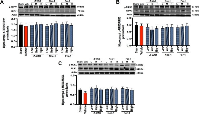
- Fig. 5 The effects of cell death inhibitors on hippocampal necroptosis signaling pathways. A p-RIPK1/RIPK1 protein expression; B p-RIPK3/RIPK3 protein expression; C p-MLKL/MLKL protein expression. Data are shown as mean +- SEM. n = 6/group. Veh: cardiac I/R rats treated with vehicle; L: cardiac I/R rats treated with low dose of cell death inhibitor; M: cardiac I/R rats treated with medium dose of cell death inhibitor; H: cardiac I/R rats treated with high dose of cell death inhibitor. I/R ischemia/reperfusion injury, RIPK receptor interacting protein kinases, MLKL mixed lineage kinase domain-like protein.