Explore
 Validate
 Learn
Learn Western blot
Antibody data
- Antibody Data
- Antigen structure
- References [1]
- Comments [0]
- Validations
- Western blot [1]
- Immunocytochemistry [1]
- Other assay [1]
Submit
Validation data
Reference
Comment
Report error
- Product number
- PA5-115578 - Provider product page

- Provider
- Invitrogen Antibodies
- Product name
- MLKL Polyclonal Antibody
- Antibody type
- Polyclonal
- Antigen
- Synthetic peptide
- Description
- Antibody detects endogenous levels of total MLKL.
- Reactivity
- Human, Mouse, Rat
- Host
- Rabbit
- Isotype
- IgG
- Vial size
- 100 μL
- Concentration
- 1 mg/mL
- Storage
- -20°C
Submitted references RIP3 Contributes to Cardiac Hypertrophy by Influencing MLKL-Mediated Calcium Influx.
Xue H, Shi H, Zhang F, Li H, Li C, Han Q
Oxidative medicine and cellular longevity 2022;2022:5490553
Oxidative medicine and cellular longevity 2022;2022:5490553
No comments: Submit comment
Supportive validation
- Submitted by
- Invitrogen Antibodies (provider)
- Main image
- Experimental details
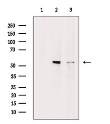
- Western blot analysis of MLKL in various samples (Lane 1: Rat liver, blocked with antigen-specific peptides, Lane 2: Rat liver, Lane 3: 293 cells (heat shock treatment)). Samples were incubated with polyclonal antibody (Product # PA5-115578).
Supportive validation
- Submitted by
- Invitrogen Antibodies (provider)
- Main image
- Experimental details
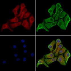
- Immunocytochemistry analysis of MLKL in HeLa cells. Samples were treated with PFA, permeabilized in 0.1% Triton X-100, blocked in 10% serum (45 min at 25°C), and incubated with polyclonal antibody (Product # PA5-115578). Secondary staining was applied with mouse anti-beta tubulin (1 hr at 37°, AlexaFluor 594 conjugated goat anti-rabbit IgG (Red), AlexaFluor 488 conjugated goat anti-mouse IgG (Green) and DAPI.

Supportive validation
- Submitted by
- Invitrogen Antibodies (provider)
- Main image
- Experimental details
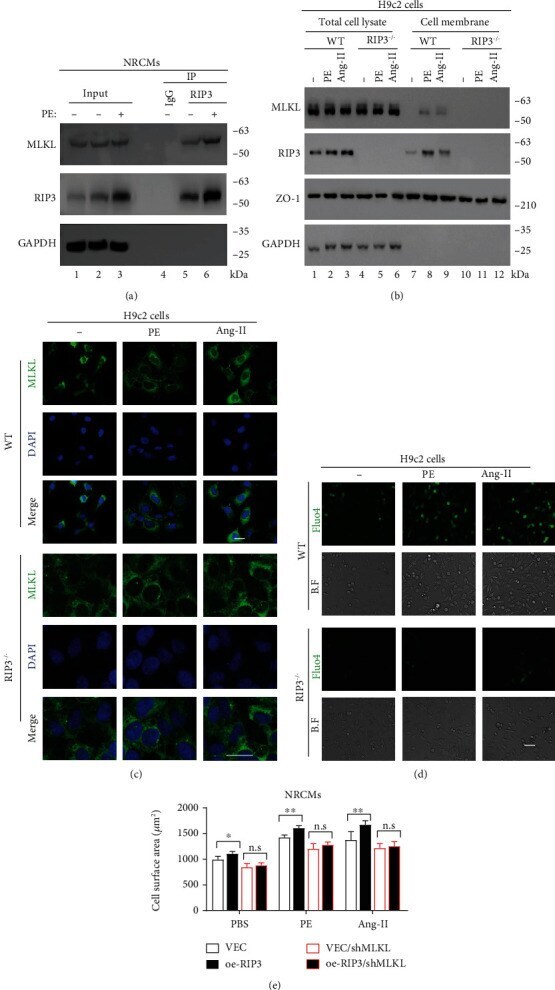
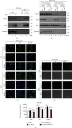
- Figure 4 RIP3 is implicated in the MLKL-mediated calcium influx. (a) Co-IP of RIP3 and MLKL in NRCMs stimulated with PE. (b) Western blot showed the cell membrane fraction assay of WT and RIP3 -/- H9c2 cells, RIP3 -/- , RIP3 knockout. WT and RIP3 -/- H9c2 cells were stimulated with Ang-II (1 mu M), PE (50 mu M), and PBS for 24 hrs. (c) Immunofluorescence of MLKL in WT and RIP3 -/- H9c2 cells stimulated with Ang-II or PE. The same cells in (b) were used. (d) Intracellular calcium concentration of H9c2 cells was determined by Fluo4 staining. The same cells in (c) were stimulated with Ang-II (1 mu M) or PE (50 mu M) for 24 hrs. (e) The surface area of the indicated NRCM cell lines. Cells were stimulated with Ang-II (1 mu M) or PE (50 mu M) for 24 hrs. VEC: infected with vector plenti; VEC/shMLKL: infected with vector/shMLKL plenti; oe-RIP3: infected with RIP3 plenti; oe-RIP3/shMLKL: infected with RIP3/shMLKL plenti. kDa: kilo-Dalton; * P < 0.05, ** P < 0.01, and *** P < 0.001.