Explore
 Validate
 Learn
Learn Western blot
Antibody data
- Antibody Data
- Antigen structure
- References [1]
- Comments [0]
- Validations
- Western blot [1]
- Immunohistochemistry [2]
Submit
Validation data
Reference
Comment
Report error
- Product number
- AF4975 - Provider product page

- Provider
- R&D Systems
- Product name
- Human 5T4 Antibody
- Antibody type
- Polyclonal
- Description
- Immunogen affinity purified. Detects human 5T4 in direct ELISAs and Western blots. In direct ELISAs and Western blots, approximately 20% cross-reactivity with recombinant mouse 5T4 is observed.
- Reactivity
- Human
- Host
- Sheep
- Conjugate
- Unconjugated
- Antigen sequence
Q13641- Isotype
- IgG
- Vial size
- 100 ug
- Concentration
- LYOPH
- Storage
- Use a manual defrost freezer and avoid repeated freeze-thaw cycles. 12 months from date of receipt, -20 to -70 °C as supplied. 1 month, 2 to 8 °C under sterile conditions after reconstitution. 6 months, -20 to -70 °C under sterile conditions after reconstitution.
Submitted references Detecting expression of 5T4 in CTCs and tumor samples from NSCLC patients.
Pirie-Shepherd SR, Painter C, Whalen P, Vizcarra P, Roy M, Qian J, Franks T, Coskran T, Golas J, Deng S, Zhong W, Tucker E, Marrinucci D, Gerber HP, Powell EL
PloS one 2017;12(7):e0179561
PloS one 2017;12(7):e0179561
No comments: Submit comment
Supportive validation
- Submitted by
- R&D Systems (provider)
- Main image
- Experimental details
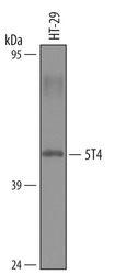
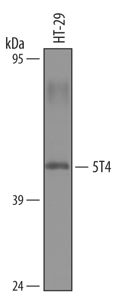
- Detection of Human 5T4 by Western Blot. Western blot shows lysates of HT-29 human colon adenocarcinoma cell line. PVDF membrane was probed with 1 µg/mL of Sheep Anti-Human 5T4 Antigen Affinity-purified Polyclonal Antibody (Catalog # AF4975) followed by HRP-conjugated Anti-Sheep IgG Secondary Antibody (Catalog # HAF016). A specific band was detected for 5T4 at approximately 50 kDa (as indicated). This experiment was conducted under reducing conditions and using Immunoblot Buffer Group 8.
Supportive validation
- Submitted by
- R&D Systems (provider)
- Main image

- Experimental details
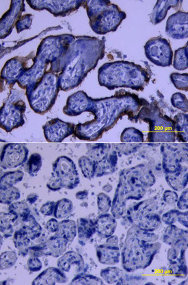
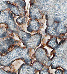
- 5T4 in Human Placenta. 5T4 was detected in immersion fixed paraffin-embedded sections of human placenta using 10 µg/mL Sheep Anti-Human 5T4 Antigen Affinity-purified Polyclonal Antibody (Catalog # AF4975) overnight at 4 °C. Before incubation with the primary antibody tissue was subjected to heat-induced epitope retrieval using Antigen Retrieval Reagent-Basic (Catalog # CTS013). Tissue was stained with the Anti-Sheep HRP-DAB Cell & Tissue Staining Kit (brown; Catalog # CTS019) and counterstained with hematoxylin (blue). View our protocol for Chromogenic IHC Staining of Paraffin-embedded Tissue Sections.
- Submitted by
- R&D Systems (provider)
- Main image

- Experimental details
- 5T4 in Human Placenta. 5T4 was detected in immersion fixed paraffin-embedded sections of human placenta using Sheep Anti-Human 5T4 Antigen Affinity-purified Polyclonal Antibody (Catalog # AF4975) at 10 µg/mL overnight at 4 °C. Tissue was stained using the Anti-Sheep HRP-DAB Cell & Tissue Staining Kit (brown; Catalog # CTS019) and counterstained with hematoxylin (blue). Lower panel shows a lack of labeling if primary antibodies are omitted and tissue is stained only with secondary antibody followed by incubation with detection reagents. View our protocol for Chromogenic IHC Staining of Paraffin-embedded Tissue Sections.