Explore
 Validate
 Learn
Learn Western blot
Antibody data
- Antibody Data
- Antigen structure
- References [0]
- Comments [0]
- Validations
- Western blot [1]
- Immunocytochemistry [3]
- Immunohistochemistry [1]
Submit
Validation data
Reference
Comment
Report error
- Product number
- PA5-35213 - Provider product page

- Provider
- Invitrogen Antibodies
- Product name
- UVRAG Polyclonal Antibody
- Antibody type
- Polyclonal
- Antigen
- Synthetic peptide
- Reactivity
- Human, Mouse
- Host
- Rabbit
- Isotype
- IgG
- Vial size
- 400 µL
- Concentration
- 2 mg/mL
- Storage
- -20° C, Avoid Freeze/Thaw Cycles
No comments: Submit comment
Supportive validation
- Submitted by
- Invitrogen Antibodies (provider)
- Main image
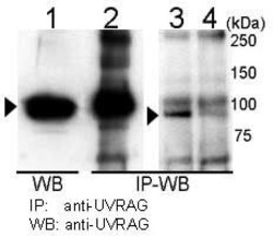
- Experimental details
- Immunoprecipitation and Western blot analysis of UVRAG in 293T cells using a UVRAG polyclonal antibody (Product # PA5-35213). UVRAG was immunoprecipitated (lane 2) and detected in 293T cells transiently transfected with mouse UVRAG (lane 1). Detection of endogenous UVRAG is shown in 293T cells (lane 3) but is reduced by UVRAG siRNA transfection (lane 4).

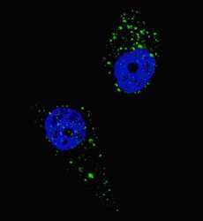
Supportive validation
- Submitted by
- Invitrogen Antibodies (provider)
- Main image

- Experimental details
- Immunofluorescent analysis of UVRAG showing staining in the cytoplasm of U-251 cells. U251 cells were treated with Chloroquine (50uM, 16h), fixed with 4% PFA (20 min) and permeabilized with Triton X-100 (0.2%, 30 min). Cells were probed with a UVRAG polyclonal antibody (Product # PA5-35213) (1:100, 2h at room temperature) followed by detection using a fluorescent conjugated secondary antibody (green) (1:1000, 1h). Nuclei were stained with Hoechst 33342 (blue) (10 µg/mL, 5 min).

- Submitted by
- Invitrogen Antibodies (provider)
- Main image

- Experimental details
- Immunofluorescent analysis of UVRAG in methanol-fixed Hela cells using a UVRAG polyclonal antibody (Product # PA5-35213).

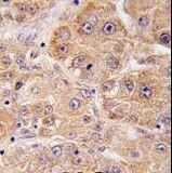
Supportive validation
- Submitted by
- Invitrogen Antibodies (provider)
- Main image

- Experimental details
- Immunohistochemistry analysis of UVRAG in formalin-fixed, paraffin-embedded human hepatocarcinoma tissue using a UVRAG polyclonal antibody (Product # PA5-35213) followed by DAB staining.