Explore
 Validate
 Learn
Learn Immunocytochemistry
 Immunohistochemistry
Antibody data
- Antibody Data
- Antigen structure
- References [4]
- Comments [0]
- Validations
- Immunocytochemistry [1]
- Chromatin Immunoprecipitation [1]
Submit
Validation data
Reference
Comment
Report error
- Product number
- HPA011165 - Provider product page

- Provider
- Atlas Antibodies
- Proper citation
- Atlas Antibodies Cat#HPA011165, RRID:AB_1856900
- Product name
- Anti-SIRT2
- Antibody type
- Polyclonal
- Description
- Polyclonal Antibody against Human SIRT2, Gene description: sirtuin 2, Alternative Gene Names: SIR2L, Validated applications: IHC, ICC, ChIP, Uniprot ID: Q8IXJ6, Storage: Store at +4°C for short term storage. Long time storage is recommended at -20°C.
- Reactivity
- Human
- Host
- Rabbit
- Conjugate
- Unconjugated
- Isotype
- IgG
- Vial size
- 100 µl
- Concentration
- 0.3 mg/ml
- Storage
- Store at +4°C for short term storage. Long time storage is recommended at -20°C.
- Handling
- The antibody solution should be gently mixed before use.
Submitted references The SIRT2/cMYC Pathway Inhibits Peroxidation-Related Apoptosis In Cholangiocarcinoma Through Metabolic Reprogramming
Expression profile of SIRT2 in human melanoma and implications for sirtuin-based chemotherapy
Reduced expression of SIRT2 in serous ovarian carcinoma promotes cell proliferation through disinhibition of CDK4 expression
A chromatin modifier genetic screen identifies SIRT2 as a modulator of response to targeted therapies through the regulation of MEK kinase activity
Xu L, Wang L, Zhou L, Dorfman R, Pan Y, Tang D, Wang Y, Yin Y, Jiang C, Zou X, Wu J, Zhang M
Neoplasia 2019;21(5):429-441
Neoplasia 2019;21(5):429-441
Expression profile of SIRT2 in human melanoma and implications for sirtuin-based chemotherapy
Wilking-Busch M, Ndiaye M, Huang W, Ahmad N
Cell Cycle 2017;16(6):574-577
Cell Cycle 2017;16(6):574-577
Reduced expression of SIRT2 in serous ovarian carcinoma promotes cell proliferation through disinhibition of CDK4 expression
Du Y, Wu J, Zhang H, Li S, Sun H
Molecular Medicine Reports 2017;15(4):1638-1646
Molecular Medicine Reports 2017;15(4):1638-1646
A chromatin modifier genetic screen identifies SIRT2 as a modulator of response to targeted therapies through the regulation of MEK kinase activity
Bajpe P, Prahallad A, Horlings H, Nagtegaal I, Beijersbergen R, Bernards R
Oncogene 2014;34(4):531-536
Oncogene 2014;34(4):531-536
No comments: Submit comment
Supportive validation
- Submitted by
- Atlas Antibodies (provider)
- Main image
- Experimental details
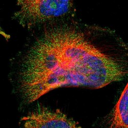
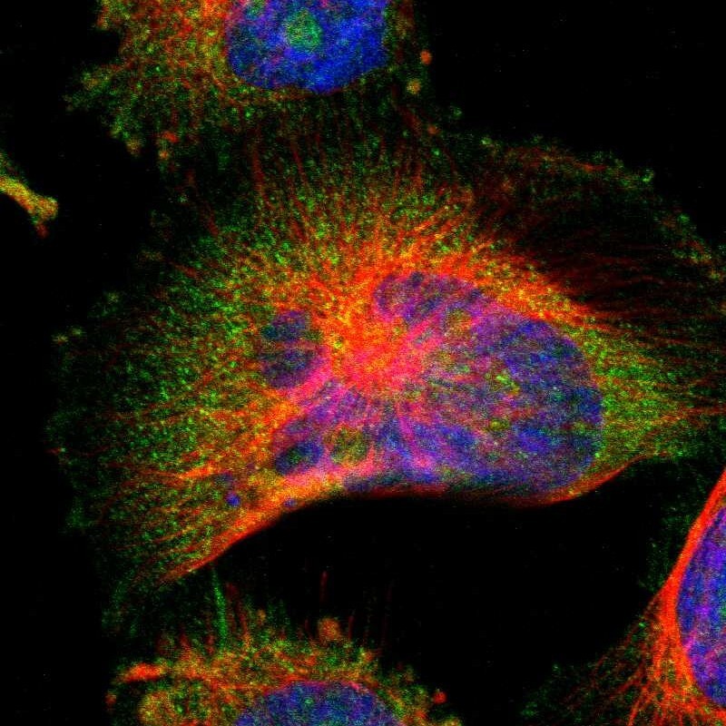
- Immunofluorescent staining of human cell line U-251 MG shows localization to nucleoli, plasma membrane & cytosol.
- Sample type
- Human
Supportive validation
- Submitted by
- Atlas Antibodies (provider)
- Main image

- Experimental details
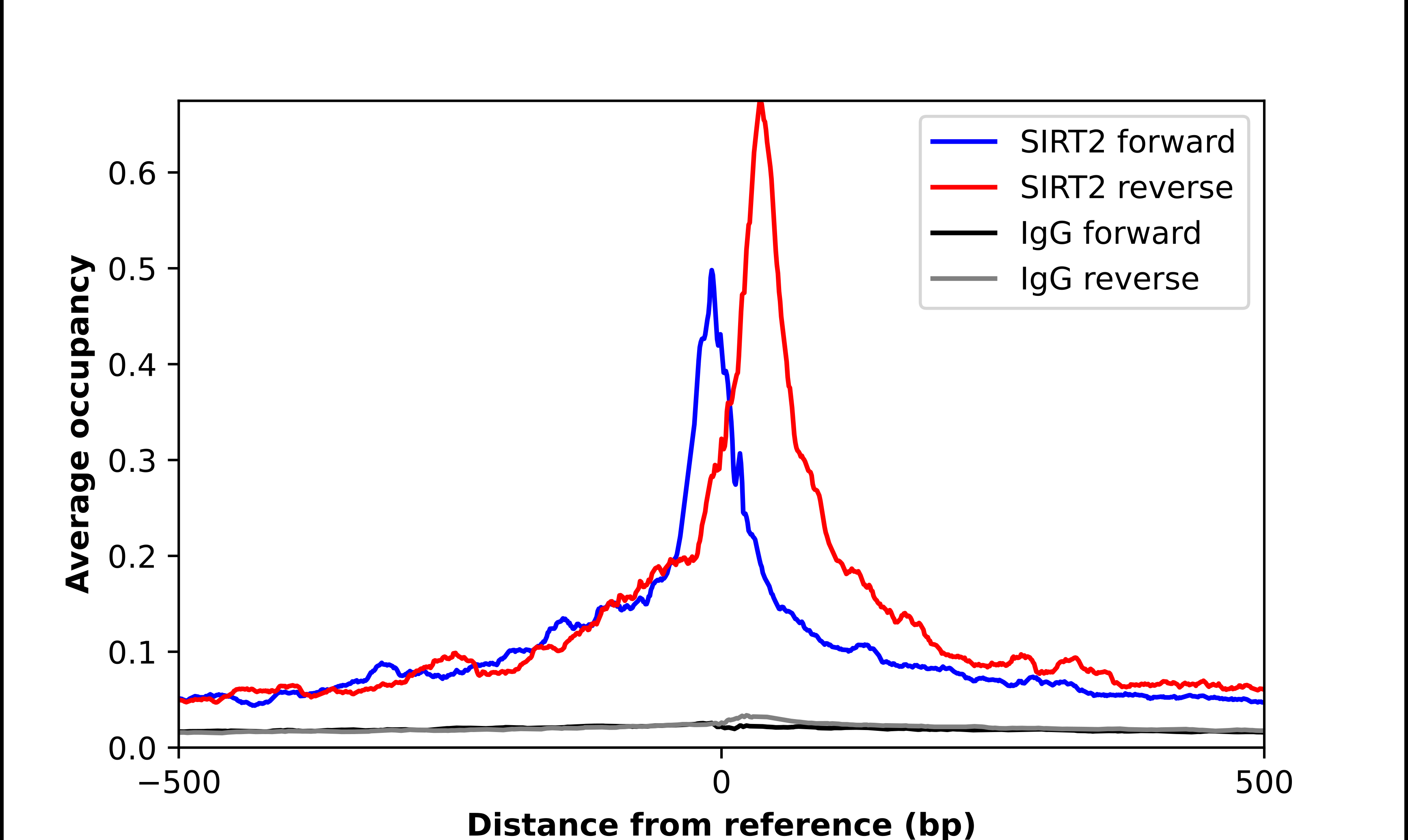
- ChIP-Exo-Seq composite graph for Anti-SIRT2 (HPA011165, Lot 000048978) tested in K562 cells. Strand-specific reads (blue: forward, red: reverse) and IgG controls (black: forward, grey: reverse) are plotted against the distance from a composite set of reference binding sites. The antibody exhibits robust target enrichment compared to a non-specific IgG control and precisely reveals its structural organization around the binding site. Data generated by Prof. B. F. Pugh´s Lab at Cornell University.