Explore
 Validate
 Learn
Learn Western blot
 Immunocytochemistry
Antibody data
- Antibody Data
- Antigen structure
- References [2]
- Comments [0]
- Validations
- Immunocytochemistry [1]
- Other assay [1]
Submit
Validation data
Reference
Comment
Report error
- Product number
- 711020 - Provider product page

- Provider
- Invitrogen Antibodies
- Product name
- GLT-1 Recombinant Superclonal™ Antibody (9 HCLC)
- Antibody type
- Other
- Antigen
- Synthetic peptide
- Description
- This antibody is predicted to react with Monkey, Sheep and Cat. Recombinant rabbit Superclonal™ antibodies are unique offerings from Thermo Fisher Scientific. They are comprised of a selection of multiple different recombinant monoclonal antibodies, providing the best of both worlds - the sensitivity of polyclonal antibodies with the specificity of monoclonal antibodies - all delivered with the consistency only found in a recombinant antibody. While functionally the same as a polyclonal antibody - recognizing multiple epitope sites on the target and producing higher detection sensitivity for low abundance targets - a recombinant rabbit Superclonal™ antibody has a known mixture of light and heavy chains. The exact population can be produced in every lot, circumventing the biological variability typically associated with polyclonal antibody production. Note: Formerly called “Recombinant polyclonal antibody”, this product is now rebranded as “Recombinant Superclonal™ antibody”. The physical product and the performance remain unchanged.
- Reactivity
- Human, Mouse, Rat
- Host
- Rabbit
- Isotype
- IgG
- Antibody clone number
- 9 HCLC
- Vial size
- 100 μg
- Concentration
- 0.5 mg/mL
- Storage
- Store at 4°C short term. For long term storage, store at -20°C, avoiding freeze/thaw cycles.
Submitted references Semaphorin 4D is upregulated in neurons of diseased brains and triggers astrocyte reactivity.
ALS-associated genes in SCA2 mouse spinal cord transcriptomes.
Evans EE, Mishra V, Mallow C, Gersz EM, Balch L, Howell A, Reilly C, Smith ES, Fisher TL, Zauderer M
Journal of neuroinflammation 2022 Aug 6;19(1):200
Journal of neuroinflammation 2022 Aug 6;19(1):200
ALS-associated genes in SCA2 mouse spinal cord transcriptomes.
Scoles DR, Dansithong W, Pflieger LT, Paul S, Gandelman M, Figueroa KP, Rigo F, Bennett CF, Pulst SM
Human molecular genetics 2020 Jun 27;29(10):1658-1672
Human molecular genetics 2020 Jun 27;29(10):1658-1672
No comments: Submit comment
Supportive validation
- Submitted by
- Invitrogen Antibodies (provider)
- Main image
- Experimental details
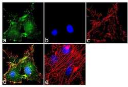
- Immunofluorescence was performed on fixed and permeabilized U-87 MG cells for detection of endogenous EAAT2 using Anti-EAAT2 Recombinant Rabbit Superclonal™ Antibody (Product # 711020, 2 µg/mL) and labeled with Goat anti-Rabbit IgG (Heavy Chain) Superclonal™ Secondary Antibody, Alexa Fluor® 488 conjugate (Product # A27034, 1:2000). Panel a) shows representative cells that were stained for detection and localization of EAAT2 protein (green), Panel b) is stained for nuclei (blue) using SlowFade® Gold Antifade Mountant with DAPI (Product # S36938). Panel c) represents cytoskeletal F-actin staining using Alexa Fluor® 555 Rhodamine Phalloidin (Product # R415, 1:300). Panel d) is a composite image of Panels a, b and c clearly demonstrating membrane localization of EAAT2. Panel e) represents control cells with no primary antibody to assess background.
Supportive validation
- Submitted by
- Invitrogen Antibodies (provider)
- Main image

- Experimental details
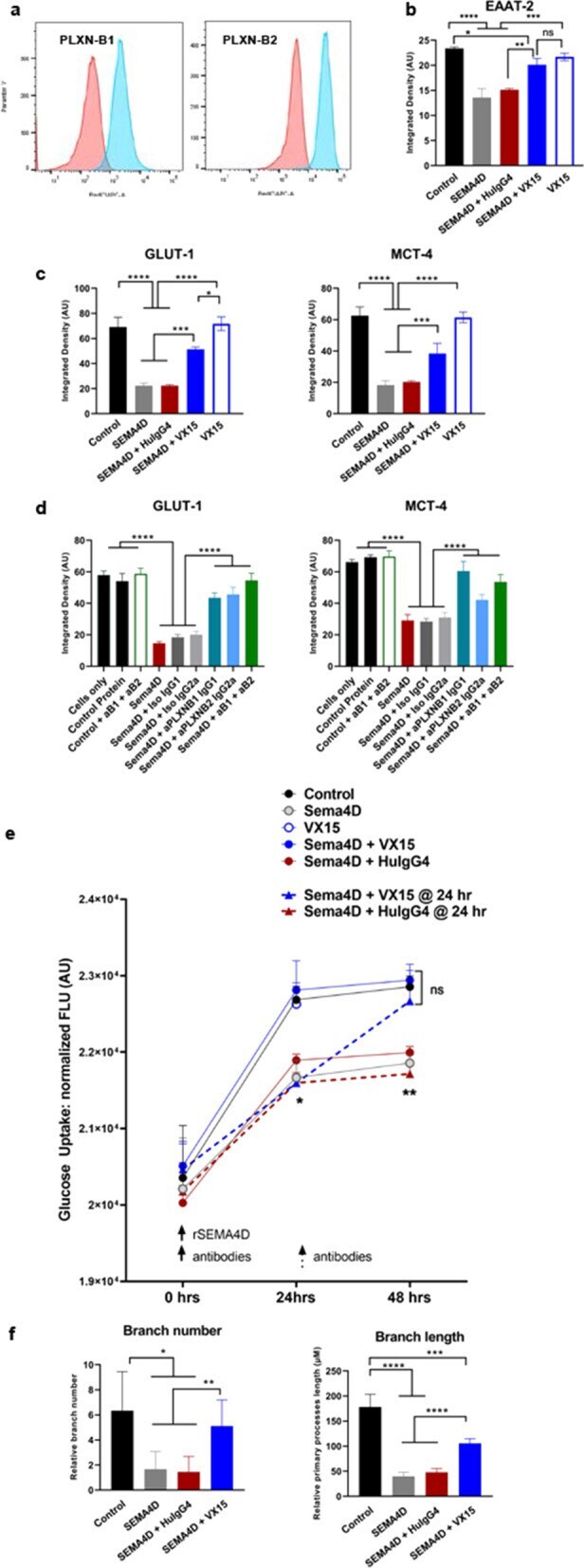
- Fig. 5 SEMA4D triggers receptor-mediated astrocyte reactivity, including changes in astrocyte morphology, expression of key transporters for glutamate recycling and energy metabolism and impairs astrocyte function of glucose uptake. a Primary human astrocyte cultures were stained for PLXN receptors (blue), compared to isotype control antibodies (red). Antibody blocking effect was determined by incubation with rSEMA4D or control protein, in presence/absence of anti-SEMA4D antibody/VX15 (human IgG4) or isotype-matched control antibodies for 48 h. Cultures were stained for b EAAT-2, and c GLUT-1 and MCT-4 transporters. d In a separate experiment, blocking of receptors was assessed using anti-PLXNB1 (mouse IgG1) and/or anti-PLXNB2 (mouse IgG2a) or isotype-matched control antibodies and the same conditions as above. Quantification is shown as; mean + SEM of replicates for each treatment. e Glucose uptake was measured in human astrocyte cultures treated as above. rSEMA4D was added at time 0 and antibodies were added at t = 0 (solid lines and circles) for inhibition or t = 24 h (dotted lines and triangles) to evaluate reversal of activity. Quantification for each condition is shown as average + SEM from 3 wells/condition/timepoint. f Morphologic changes showing length and number of primary processes. For b to f , multivariate regression analysis was performed to determine the effect treatment conditions and dependent variables with treatment type. The significance levels are reporte