Explore
 Validate
 Learn
Learn Western blot
 Immunoprecipitation
Antibody data
- Antibody Data
- Antigen structure
- References [3]
- Comments [0]
- Validations
- Western blot [1]
- Immunohistochemistry [1]
Submit
Validation data
Reference
Comment
Report error
- Product number
- AF3300 - Provider product page

- Provider
- R&D Systems
- Product name
- Mouse Meprin beta Subunit/MEP1B Antibody
- Antibody type
- Polyclonal
- Description
- Immunogen affinity purified. Detects mouse Meprin beta Subunit/MEP1B in direct ELISAs and Western blots. In Western blots, approximately 40% cross-reactivity with recombinant human MEP1B is observed.
- Reactivity
- Mouse
- Host
- Goat
- Conjugate
- Unconjugated
- Antigen sequence
Q61847- Isotype
- IgG
- Vial size
- 100 ug
- Concentration
- LYOPH
- Storage
- Use a manual defrost freezer and avoid repeated freeze-thaw cycles. 12 months from date of receipt, -20 to -70 °C as supplied. 1 month, 2 to 8 °C under sterile conditions after reconstitution. 6 months, -20 to -70 °C under sterile conditions after reconstitution.
Submitted references Meprin β contributes to collagen deposition in lung fibrosis.
ADAM10 is the major sheddase responsible for the release of membrane-associated meprin A.
Microbial-induced meprin β cleavage in MUC2 mucin and a functional CFTR channel are required to release anchored small intestinal mucus.
Biasin V, Wygrecka M, Marsh LM, Becker-Pauly C, Brcic L, Ghanim B, Klepetko W, Olschewski A, Kwapiszewska G
Scientific reports 2017 Jan 6;7:39969
Scientific reports 2017 Jan 6;7:39969
ADAM10 is the major sheddase responsible for the release of membrane-associated meprin A.
Herzog C, Haun RS, Ludwig A, Shah SV, Kaushal GP
The Journal of biological chemistry 2014 May 9;289(19):13308-22
The Journal of biological chemistry 2014 May 9;289(19):13308-22
Microbial-induced meprin β cleavage in MUC2 mucin and a functional CFTR channel are required to release anchored small intestinal mucus.
Schütte A, Ermund A, Becker-Pauly C, Johansson ME, Rodriguez-Pineiro AM, Bäckhed F, Müller S, Lottaz D, Bond JS, Hansson GC
Proceedings of the National Academy of Sciences of the United States of America 2014 Aug 26;111(34):12396-401
Proceedings of the National Academy of Sciences of the United States of America 2014 Aug 26;111(34):12396-401
No comments: Submit comment
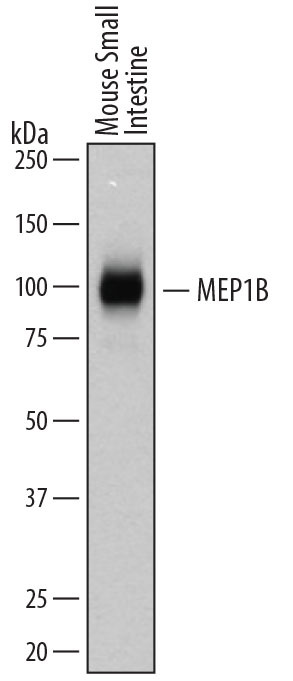
Supportive validation
- Submitted by
- R&D Systems (provider)
- Main image
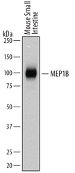
- Experimental details
- Detection of Mouse Meprin beta Subunit/MEP1B by Western Blot. Western blot shows lysates of mouse small intestine tissue. PVDF membrane was probed with 0.1 µg/mL of Goat Anti-Mouse Meprin beta Subunit/MEP1B Antigen Affinity-purified Polyclonal Antibody (Catalog # AF3300) followed by HRP-conjugated Anti-Goat IgG Secondary Antibody (Catalog # HAF109). A specific band was detected for Meprin beta Subunit/MEP1B at approximately 97 kDa (as indicated). This experiment was conducted under reducing conditions and using Immunoblot Buffer Group 5.
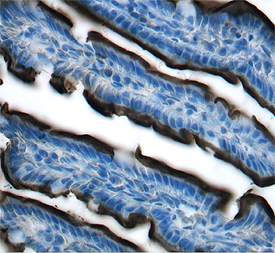
Supportive validation
- Submitted by
- R&D Systems (provider)
- Main image

- Experimental details
- Meprin beta Subunit/MEP1B in Mouse Intestine. Meprin beta Subunit/MEP1B was detected in perfusion fixed frozen sections of mouse intestine using 1 µg/mL Goat Anti-Mouse Meprin beta Subunit/MEP1B Antigen Affinity-purified Polyclonal Antibody (Catalog # AF3300) overnight at 4 °C. Tissue was stained with the Anti-Goat HRP-DAB Cell & Tissue Staining Kit (brown; Catalog # CTS008) and counterstained with hematoxylin (blue). Specific labeling was localized to the brush border of intestinal villi.View our protocol for Chromogenic IHC Staining of Frozen Tissue Sections.