 Explore
 Validate
 Learn
Learn Western blot
Antibody data
- Antibody Data
- Antigen structure
- References [1]
- Comments [0]
- Validations
- Western blot [1]
- Immunocytochemistry [1]
- Immunohistochemistry [1]
Submit
Validation data
Reference
Comment
Report error
- Product number
- AF7964-100 - Provider product page

- Provider
- R&D Systems
- Product name
- Human RNF4 Antibody
- Antibody type
- Polyclonal
- Description
- Antigen Affinity-purified. Detects human RNF4 in direct ELISAs and Western blots.
- Reactivity
- Human
- Host
- Goat
- Conjugate
- Unconjugated
- Antigen sequence
P78317- Isotype
- IgG
- Vial size
- 100 ug
- Storage
- Use a manual defrost freezer and avoid repeated freeze-thaw cycles. 12 months from date of receipt, -20 to -70 °C as supplied. 1 month, 2 to 8 °C under sterile conditions after reconstitution. 6 months, -20 to -70 °C under sterile conditions after reconstitution.
Submitted references Inhibiting ubiquitination causes an accumulation of SUMOylated newly synthesized nuclear proteins at PML bodies.
Sha Z, Blyszcz T, González-Prieto R, Vertegaal ACO, Goldberg AL
The Journal of biological chemistry 2019 Oct 18;294(42):15218-15234
The Journal of biological chemistry 2019 Oct 18;294(42):15218-15234
No comments: Submit comment
Supportive validation
- Submitted by
- R&D Systems (provider)
- Main image
- Experimental details
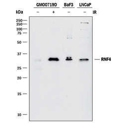
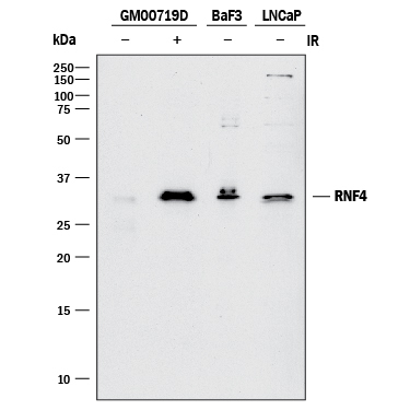
- Detection of Human RNF4 by Western Blot. Western blot shows lysates of GM00719D human ataxia telangiectasia cell line either mock-treated (-) or exposed (+) to 10 Gy ionizing radiation (IR) and harvested after 1 hour, BaF3 mouse pro-B cell line, and LNCaP human prostate cancer cell line. PVDF membrane was probed with 1 µg/mL of Goat Anti-Human RNF4 Antigen Affinity-purified Polyclonal Antibody (Catalog # AF7964) followed by HRP-conjugated Anti-Goat IgG Secondary Antibody (Catalog # HAF017). A specific band was detected for RNF4 at approximately 34 kDa (as indicated). This experiment was conducted under reducing conditions and using Immunoblot Buffer Group 1.
Supportive validation
- Submitted by
- R&D Systems (provider)
- Main image

- Experimental details
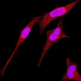
- RNF4 in HeLa Human Cell Line. RNF4 was detected in immersion fixed HeLa human cervical epithelial carcinoma cell line using Goat Anti-Human RNF4 Antigen Affinity-purified Polyclonal Antibody (Catalog # AF7964) at 1.7 µg/mL for 3 hours at room temperature. Cells were stained using the NorthernLights™ 557-conjugated Anti-Goat IgG Secondary Antibody (red; Catalog # NL001) and counterstained with DAPI (blue). Specific staining was localized to nuclei and cytoplasm. View our protocol for Fluorescent ICC Staining of Cells on Coverslips.
Supportive validation
- Submitted by
- R&D Systems (provider)
- Main image
- Experimental details
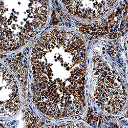
- RNF4 in Human Testis. RNF4 was detected in immersion fixed paraffin-embedded sections of human testis using Goat Anti-Human RNF4 Antigen Affinity-purified Polyclonal Antibody (Catalog # AF7964) at 10 µg/mL overnight at 4 °C. Tissue was stained using the Anti-Goat HRP-DAB Cell & Tissue Staining Kit (brown; Catalog # CTS008) and counterstained with hematoxylin (blue). Specific staining was localized to nuclei and cytoplasm. View our protocol for Chromogenic IHC Staining of Paraffin-embedded Tissue Sections.
