 Explore
 Validate
 Learn
Learn Western blot
Antibody data
- Antibody Data
- Antigen structure
- References [0]
- Comments [0]
- Validations
- Western blot [1]
- Immunohistochemistry [1]
- Flow cytometry [1]
Submit
Validation data
Reference
Comment
Report error
- Product number
- AF4234 - Provider product page

- Provider
- R&D Systems
- Product name
- Human PSMA/FOLH1/NAALADase I Antibody
- Antibody type
- Polyclonal
- Description
- Immunogen affinity purified. Detects human PSMA/FOLH1/NAALADase I in direct ELISAs and Western blots. In direct ELISAs and Western blots, less than 5% cross-reactivity with recombinant mouse NAALADase II is observed.
- Reactivity
- Human
- Host
- Sheep
- Conjugate
- Unconjugated
- Antigen sequence
Q04609- Isotype
- IgG
- Vial size
- 100 ug
- Concentration
- LYOPH
- Storage
- Use a manual defrost freezer and avoid repeated freeze-thaw cycles. 12 months from date of receipt, -20 to -70 °C as supplied. 1 month, 2 to 8 °C under sterile conditions after reconstitution. 6 months, -20 to -70 °C under sterile conditions after reconstitution.
No comments: Submit comment
Supportive validation
- Submitted by
- R&D Systems (provider)
- Main image

- Experimental details
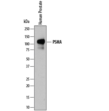
- Detection of Human PSMA/FOLH1/ NAALADase I by Western Blot. Western blot shows lysates of human prostate tissue. PVDF membrane was probed with 0.2 µg/mL of Sheep Anti-Human PSMA/FOLH1/ NAALADase I Antigen Affinity-purified Poly-clonal Antibody (Catalog # AF4234) followed by HRP-conjugated Anti-Sheep IgG Secondary Antibody (Catalog # HAF016). A specific band was detected for PSMA/FOLH1/NAALADase I at approximately 120 kDa (as indicated). This experiment was conducted under reducing conditions and using Immunoblot Buffer Group 1.
Supportive validation
- Submitted by
- R&D Systems (provider)
- Main image
- Experimental details
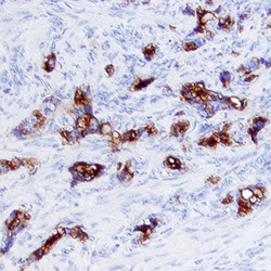
- PSMA/FOLH1/NAALADase I in Human Prostate Cancer Tissue. PSMA/FOLH1/NAALADase I was detected in immersion fixed paraffin-embedded sections of human prostate cancer tissue using Sheep Anti-Human PSMA/FOLH1/NAALADase I Antigen Affinity-purified Poly-clonal Antibody (Catalog # AF4234) at 5 µg/mL overnight at 4 °C. Tissue was stained using the Anti-Sheep HRP-DAB Cell & Tissue Staining Kit (brown; Catalog # CTS019) and counter-stained with hematoxylin (blue). Specific labeling was localized to the plasma membrane and extracellular space. View our protocol for Chromogenic IHC Staining of Paraffin-embedded Tissue Sections.

Supportive validation
- Submitted by
- R&D Systems (provider)
- Main image
- Experimental details
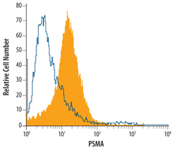
- Detection of PSMA/FOLH1/ NAALADase I in LnCAP Human Cell Line by Flow Cytometry. LnCAP human prostate cancer cell line was stained with Human PSMA/FOLH1/NAALADase I Antigen Affinity-purified Polyclonal Antibody (Catalog # AF4234, filled histo-gram) or control antibody (Catalog # 5-001-A, open histo-gram), followed by Northern-Lights™ 557-conjugated Anti-Sheep IgG Secondary Antibody (Catalog # NL010).
