Explore
 Validate
 Learn
Learn ELISA
ELISA Immunocytochemistry
Antibody data
- Antibody Data
- Antigen structure
- References [0]
- Comments [0]
- Validations
- Immunocytochemistry [2]
- Flow cytometry [1]
Submit
Validation data
Reference
Comment
Report error
- Product number
- MA5-48336 - Provider product page

- Provider
- Invitrogen Antibodies
- Product name
- PRDM9 Chimeric Recombinant Rabbit Monoclonal Antibody (RAB-C370)
- Antibody type
- Monoclonal
- Antigen
- Recombinant full-length protein
- Description
- Specificity: This antibody recognizes PRDM9 (Histone-lysine N-methyltransferase PRDM9). It binds to a folded domain, amino acids 195-385. PRDM9 is a histone methyltransferase that plays a key role in meiotic prophase by determining hotspot localization thereby promoting meiotic recombination.
- Reactivity
- Human
- Host
- Rabbit
- Isotype
- IgG
- Antibody clone number
- RAB-C370
- Vial size
- 200 μg
- Concentration
- 1 mg/mL
- Storage
- Store at 4°C short term. For long term storage, store at -20°C, avoiding freeze/thaw cycles.
No comments: Submit comment
Supportive validation
- Submitted by
- Invitrogen Antibodies (provider)
- Main image
- Experimental details
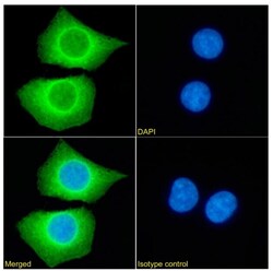
- Immunocytochemistry/Immunofluorescence analysis of PRDM9 in HEK293 cells using PRDM9 Chimeric Monoclonal Antibody (Product # MA5-48336). Cells were fixed with paraformaldehyde, permeabilized with 0.15% Triton and stained with PRDM9 Chimeric Monoclonal Antibody at a dilution of 1:100 for 1h followed by Alexa Fluor 488 secondary antibody (1:1,000 dilution), showing cytoplasmic staining. The nuclear stain is DAPI (blue). Panels show from left-right, top-bottom PRDM9 Chimeric Monoclonal Antibody, DAPI, merged channels and an isotype control. The isotype control was a Rabbit IgG Chimeric Monoclonal Antibody (Product # MA5-47825) followed by staining with Alexa Fluor 488 secondary antibody.

- Submitted by
- Invitrogen Antibodies (provider)
- Main image
- Experimental details
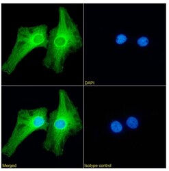
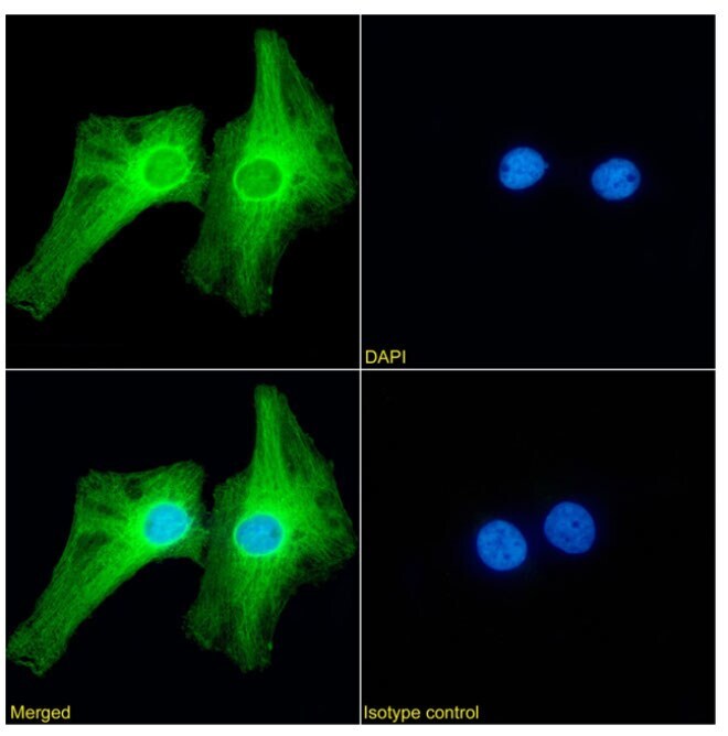
- Immunocytochemistry/Immunofluorescence analysis of PRDM9 in HeLa cells using PRDM9 Chimeric Monoclonal Antibody (Product # MA5-48336). Cells were fixed with paraformaldehyde, permeabilized with 0.15% Triton and stained with PRDM9 Chimeric Monoclonal Antibody at a dilution of 1:100 for 1h followed by Alexa Fluor 488 secondary antibody (1:1,500 dilution), showing cytoplasmic staining. The nuclear stain is DAPI (blue). Panels show from left-right, top-bottom PRDM9 Chimeric Monoclonal Antibody, DAPI, merged channels and an isotype control. The isotype control was a Rabbit IgG Chimeric Monoclonal Antibody (Product # MA5-47825) followed by staining with Alexa Fluor 488 secondary antibody.
Supportive validation
- Submitted by
- Invitrogen Antibodies (provider)
- Main image
- Experimental details
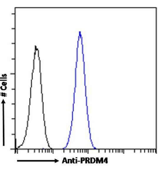
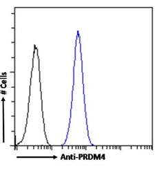
- Flow Cytometry analysis of PRDM9 in HeLa cells using PRDM9 Chimeric Monoclonal Antibody (Product # MA5-48336). Cells were fixed with paraformaldehyde, permeabilized with 0.5% triton and stained with Rabbit IgG Isotype Control (Product # MA5-47825) (black line) or PRDM9 Chimeric Monoclonal Antibody (blue line) at a dilution of 1:100 for 1h at RT. After washing, the bound antibody was detected using a goat anti-rabbit IgG AF488 antibody at a dilution of 1:1,000 and cells analyzed using a flow-cytometer.