 Explore
 Validate
 Learn
LearnNBP1-03349
antibody from Novus Biologicals
Targeting: DROSHA
Etohi2, HSA242976, RN3, RNASE3L, RNASEN
 Western blot
 Immunocytochemistry
Antibody data
- Antibody Data
- Antigen structure
- References [5]
- Comments [0]
- Validations
- Western blot [2]
- Immunoprecipitation [1]
- Immunohistochemistry [1]
Submit
Validation data
Reference
Comment
Report error
- Product number
- NBP1-03349 - Provider product page

- Provider
- Novus Biologicals
- Proper citation
- Novus Cat#NBP1-03349, RRID:AB_1520772
- Product name
- Rabbit Polyclonal Drosha Antibody
- Antibody type
- Polyclonal
- Description
- Immunogen affinity purified.
- Reactivity
- Human, Mouse
- Host
- Rabbit
- Isotype
- IgG
- Vial size
- 0.1 ml
- Concentration
- 0.2 mg/ml
- Storage
- Store at 4C. Do not freeze.
Submitted references Allele-specific loss and transcription of the miR-15a/16-1 cluster in chronic lymphocytic leukemia.
DGCR8 Acts as an Adaptor for the Exosome Complex to Degrade Double-Stranded Structured RNAs.
Hypoxia-mediated downregulation of miRNA biogenesis promotes tumour progression.
Inducible deletion of epidermal Dicer and Drosha reveals multiple functions for miRNAs in postnatal skin.
WDHD1 modulates the post-transcriptional step of the centromeric silencing pathway.
Veronese A, Pepe F, Chiacchia J, Pagotto S, Lanuti P, Veschi S, Di Marco M, D'Argenio A, Innocenti I, Vannata B, Autore F, Marchisio M, Wernicke D, Verginelli F, Leone G, Rassenti LZ, Kipps TJ, Mariani-Costantini R, Laurenti L, Croce CM, Visone R
Leukemia 2015 Jan;29(1):86-95
Leukemia 2015 Jan;29(1):86-95
DGCR8 Acts as an Adaptor for the Exosome Complex to Degrade Double-Stranded Structured RNAs.
Macias S, Cordiner RA, Gautier P, Plass M, Cáceres JF
Molecular cell 2015 Dec 17;60(6):873-85
Molecular cell 2015 Dec 17;60(6):873-85
Hypoxia-mediated downregulation of miRNA biogenesis promotes tumour progression.
Rupaimoole R, Wu SY, Pradeep S, Ivan C, Pecot CV, Gharpure KM, Nagaraja AS, Armaiz-Pena GN, McGuire M, Zand B, Dalton HJ, Filant J, Miller JB, Lu C, Sadaoui NC, Mangala LS, Taylor M, van den Beucken T, Koch E, Rodriguez-Aguayo C, Huang L, Bar-Eli M, Wouters BG, Radovich M, Ivan M, Calin GA, Zhang W, Lopez-Berestein G, Sood AK
Nature communications 2014 Oct 29;5:5202
Nature communications 2014 Oct 29;5:5202
Inducible deletion of epidermal Dicer and Drosha reveals multiple functions for miRNAs in postnatal skin.
Teta M, Choi YS, Okegbe T, Wong G, Tam OH, Chong MM, Seykora JT, Nagy A, Littman DR, Andl T, Millar SE
Development (Cambridge, England) 2012 Apr;139(8):1405-16
Development (Cambridge, England) 2012 Apr;139(8):1405-16
WDHD1 modulates the post-transcriptional step of the centromeric silencing pathway.
Hsieh CL, Lin CL, Liu H, Chang YJ, Shih CJ, Zhong CZ, Lee SC, Tan BC
Nucleic acids research 2011 May;39(10):4048-62
Nucleic acids research 2011 May;39(10):4048-62
No comments: Submit comment
Supportive validation
- Submitted by
- Novus Biologicals (provider)
- Main image

- Experimental details
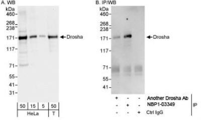
- Western Blot: Drosha Antibody [NBP1-03349] - Whole cell lysate from HeLa (5, 15 and 50 ug for WB; 1 mg for IP, 20% of IP loaded) and 293T (T; 50 ug) cells. NBP1-03349 used for WB at 0.04 ug/ml (A) and 0.1 ug/ml (B) and used for IP at 3 ug/mg lysate.
- Submitted by
- Novus Biologicals (provider)
- Main image
- Experimental details
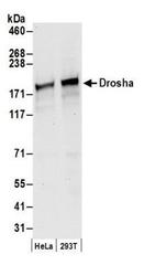
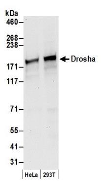
- Western Blot: Drosha Antibody [NBP1-03349] - Detection of Human Drosha by Western Blot. Samples: Whole cell lysate (50 ug) from HeLa and 293T cells prepared using NETN lysis buffer. Antibody: Affinity purified rabbit anti-Drosha antibody NBP1-03349 used for WB at 0.1 ug/ml. Detection: Chemiluminescence with an exposure time of 10 seconds.
Supportive validation
- Submitted by
- Novus Biologicals (provider)
- Main image
- Experimental details
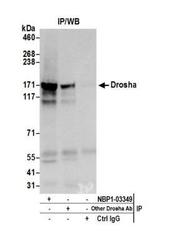
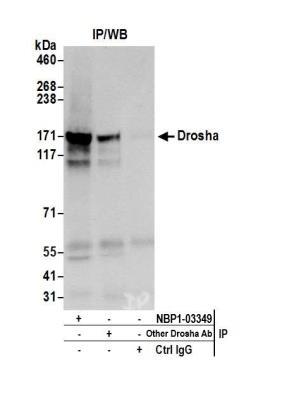
- Immunoprecipitation: Drosha Antibody [NBP1-03349] - Detection of human Drosha by western blot of immunoprecipitates. Samples: Whole cell lysate (0.5 or 1.0 mg per IP reaction; 20% of IP loaded) from HeLa cells prepared using NETN lysis buffer. Antibodies: Affinity purified rabbit anti-Drosha antibody NBP1-03349 used for IP at 6 ug per reaction. Drosha was also immunoprecipitated by another rabbit anti-Drosha antibody. For blotting immunoprecipitated Drosha, NBP1-03349 was used at 1 ug/ml. Detection: Chemiluminescence with an exposure time of 10 seconds.
Supportive validation
- Submitted by
- Novus Biologicals (provider)
- Main image
- Experimental details
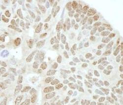
- Immunohistochemistry-Paraffin: Drosha Antibody [NBP1-03349] - FFPE human ovarian tumor. Affinity purified rabbit anti-Drosha 1:250.
