Explore
 Validate
 Learn
Learn Western blot
 Immunocytochemistry
Antibody data
- Antibody Data
- Antigen structure
- References [1]
- Comments [0]
- Validations
- Immunocytochemistry [4]
- Immunohistochemistry [4]
Submit
Validation data
Reference
Comment
Report error
- Product number
- PA5-51539 - Provider product page

- Provider
- Invitrogen Antibodies
- Product name
- UNC84B Polyclonal Antibody
- Antibody type
- Polyclonal
- Antigen
- Recombinant protein fragment
- Description
- Immunogen sequence: LKSEWQSMTQ ESFQESSVKE LRRLEDQLAG LQQELAALAL KQSSVAEEVG LLPQQIQAVR DDVESQFPAW ISQFLARGGG GRVGLLQREE MQAQLRELES KILTHVAEMQ GKSAREAAAS LSLTLQKEGV IGVTEEQVHH IVKQALQRYS E Highest antigen sequence identity to the following orthologs: Mouse - 78%, Rat - 80%.
- Reactivity
- Human
- Host
- Rabbit
- Isotype
- IgG
- Vial size
- 100 μL
- Concentration
- 0.1 mg/mL
- Storage
- Store at 4°C short term. For long term storage, store at -20°C, avoiding freeze/thaw cycles.
Submitted references Cytoplasmic control of intranuclear polarity by human cytomegalovirus.
Procter DJ, Furey C, Garza-Gongora AG, Kosak ST, Walsh D
Nature 2020 Nov;587(7832):109-114
Nature 2020 Nov;587(7832):109-114
No comments: Submit comment
Supportive validation
- Submitted by
- Invitrogen Antibodies (provider)
- Main image
- Experimental details
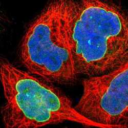
- Immunofluorescent staining of UNC84B in human cell line A-431 shows positivity in nuclear membrane. Samples were probed using an UNC84B Polyclonal Antibody (Product # PA5-51539).
- Submitted by
- Invitrogen Antibodies (provider)
- Main image

- Experimental details
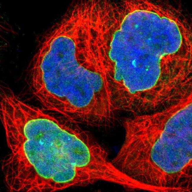
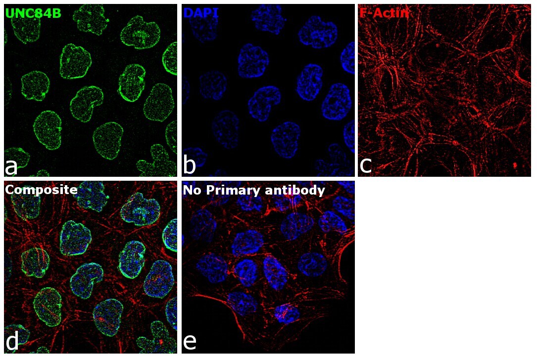
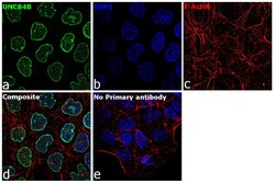
- Immunofluorescence analysis of UNC84B was performed using 70% confluent log phase A-431 cells. The cells were fixed with 4% paraformaldehyde for 10 minutes, permeabilized with 0.1% Triton™ X-100 for 15 minutes, and blocked with 2% BSA for 1 hour at room temperature. The cells were labeled with UNC84B Rabbit Polyclonal Antibody (Product # PA5-51539) at 5 µg/mL in 0.1% BSA, incubated at 4 degree Celsius overnight and then labeled with Goat anti-Rabbit IgG (H+L) Superclonal™ Secondary Antibody, Alexa Fluor® 488 conjugate (Product # A27034) at a dilution of 1:2000 for 45 minutes at room temperature (Panel a: green). Nuclei (Panel b: blue) were stained with ProLong™ Diamond Antifade Mountant with DAPI (Product # P36962). F-actin (Panel c: red) was stained with Rhodamine Phalloidin (Product # R415). Panel d represents the merged image showing Nuclear and Nuclear membrane localization. Panel e represents control cells with no primary antibody to assess background. The images were captured at 60X magnification.
- Submitted by
- Invitrogen Antibodies (provider)
- Main image

- Experimental details
- Immunofluorecent analysis of UNC84B in human cell line A-431 using UNC84B Polyclonal Antibody (Product # PA5-51539). Staining shows positivity in nuclear membrane.

- Submitted by
- Invitrogen Antibodies (provider)
- Main image
- Experimental details
- Immunofluorescence analysis of UNC84B was performed using 70% confluent log phase A-431 cells. The cells were fixed with 4% paraformaldehyde for 10 minutes, permeabilized with 0.1% Triton™ X-100 for 15 minutes, and blocked with 2% BSA for 1 hour at room temperature. The cells were labeled with UNC84B Rabbit Polyclonal Antibody (Product # PA5-51539) at 5 µg/mL in 0.1% BSA, incubated at 4 degree Celsius overnight and then labeled with Goat anti-Rabbit IgG (Heavy Chain) Superclonal™ Secondary Antibody, Alexa Fluor® 488 conjugate (Product # A27034) at a dilution of 1:2000 for 45 minutes at room temperature (Panel a: green). Nuclei (Panel b: blue) were stained with ProLong™ Diamond Antifade Mountant with DAPI (Product # P36962). F-actin (Panel c: red) was stained with Rhodamine Phalloidin (Product # R415). Panel d represents the merged image showing Nuclear and Nuclear membrane localization. Panel e represents control cells with no primary antibody to assess background. The images were captured at 60X magnification.

Supportive validation
- Submitted by
- Invitrogen Antibodies (provider)
- Main image
- Experimental details
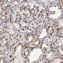
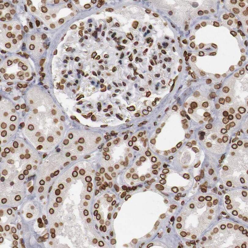
- Immunohistochemical staining of UNC84B in human kidney using an UNC84B Polyclonal Antibody (Product # PA5-51539) shows moderate to strong positivity in nuclear membrane in cells in tubules and cells in glomeruli.
- Submitted by
- Invitrogen Antibodies (provider)
- Main image

- Experimental details
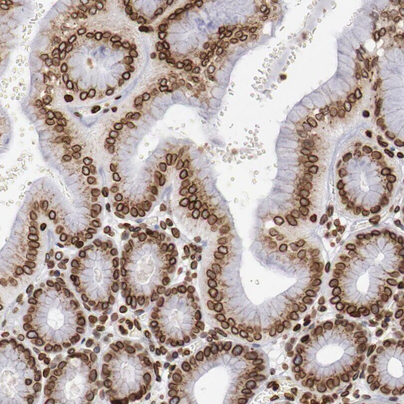
- Immunohistochemical staining of UNC84B in human stomach using an UNC84B Polyclonal Antibody (Product # PA5-51539) shows moderate to strong positivity in nuclear membrane in glandular cells.
- Submitted by
- Invitrogen Antibodies (provider)
- Main image
- Experimental details
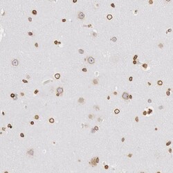
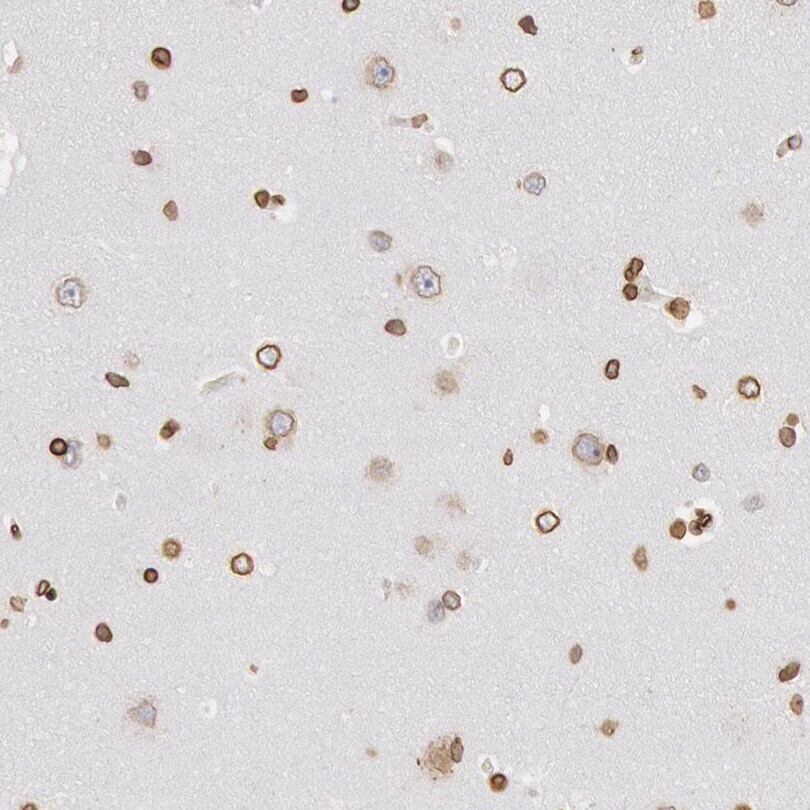
- Immunohistochemical staining of UNC84B in human cerebral cortex using an UNC84B Polyclonal Antibody (Product # PA5-51539) shows moderate to strong positivity in nuclear membrane in neuronal cells and glial cells.
- Submitted by
- Invitrogen Antibodies (provider)
- Main image
- Experimental details
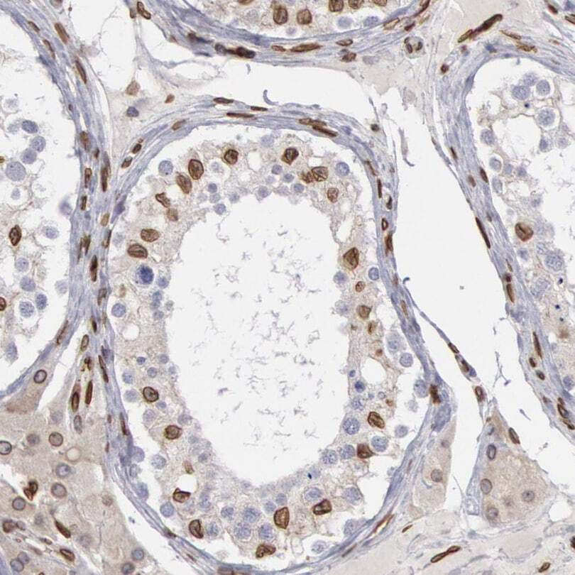
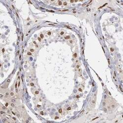
- Immunohistochemical staining of UNC84B in human testis using an UNC84B Polyclonal Antibody (Product # PA5-51539) shows moderate to strong positivity in nuclear membrane in cells in seminiferous ducts.