Explore
 Validate
 Learn
Learn Western blot
 Immunocytochemistry
 Immunohistochemistry
Antibody data
- Antibody Data
- Antigen structure
- References [16]
- Comments [0]
- Validations
- Western blot [1]
- Immunocytochemistry [1]
Submit
Validation data
Reference
Comment
Report error
- Product number
- HPA001209 - Provider product page

- Provider
- Atlas Antibodies
- Proper citation
- Atlas Antibodies Cat#HPA001209, RRID:AB_1080465
- Product name
- Anti-SUN2
- Antibody type
- Polyclonal
- Description
- Polyclonal Antibody against Human SUN2, Gene description: Sad1 and UNC84 domain containing 2, Alternative Gene Names: UNC84B, Validated applications: WB, IHC, ICC, Uniprot ID: Q9UH99, Storage: Store at +4°C for short term storage. Long time storage is recommended at -20°C.
- Reactivity
- Human
- Host
- Rabbit
- Conjugate
- Unconjugated
- Isotype
- IgG
- Vial size
- 100 µl
- Concentration
- 0.1 mg/ml
- Storage
- Store at +4°C for short term storage. Long time storage is recommended at -20°C.
- Handling
- The antibody solution should be gently mixed before use.
Submitted references Nucleocytoplasmic transport rates are regulated by cellular processes that modulate GTP availability.
The LINC complex ensures accurate centrosome positioning during prophase.
Activation of endoplasmic reticulum stress in premature aging via the inner nuclear membrane protein SUN2
SUN2 Modulates the Propagation of HSV-1
Lamin A and the LINC complex act as potential tumor suppressors in Ewing Sarcoma
Nuclear decoupling is part of a rapid protein-level cellular response to high-intensity mechanical loading
Biallelic mutations in nucleoporin NUP88 cause lethal fetal akinesia deformation sequence
TRIM11, a direct target of miR-24-3p, promotes cell proliferation and inhibits apoptosis in colon cancer
Expression of Leukemia-Associated Nup98 Fusion Proteins Generates an Aberrant Nuclear Envelope Phenotype
Nuclear envelope-associated endosomes deliver surface proteins to the nucleus
SUN2 exerts tumor suppressor functions by suppressing the Warburg effect in lung cancer
BRD4 Short Isoform Interacts with RRP1B, SIPA1 and Components of the LINC Complex at the Inner Face of the Nuclear Membrane
A mammalian KASH domain protein coupling meiotic chromosomes to the cytoskeleton
Immunofluorescence and fluorescent-protein tagging show high correlation for protein localization in mammalian cells
Samp1 is functionally associated with the LINC complex and A-type lamina networks
Scott KL, Halfmann CT, Hoefakker AD, Purkayastha P, Wang TC, Lele TP, Roux KJ
The Journal of cell biology 2024 Jul 1;223(7)
The Journal of cell biology 2024 Jul 1;223(7)
The LINC complex ensures accurate centrosome positioning during prophase.
Lima JT, Pereira AJ, Ferreira JG
Life science alliance 2024 Apr;7(4)
Life science alliance 2024 Apr;7(4)
Wiggan O, Stasevich T
2024
2024
Activation of endoplasmic reticulum stress in premature aging via the inner nuclear membrane protein SUN2
Vidak S, Serebryannyy L, Pegoraro G, Misteli T
Cell Reports 2023;42(5):112534
Cell Reports 2023;42(5):112534
SUN2 Modulates the Propagation of HSV-1
Cruz-Palomar K, Hawkins J, Vandal C, Quenneville J, Gagnon É, Lippé R, Sandri-Goldin R
Journal of Virology 2022;96(9)
Journal of Virology 2022;96(9)
Lamin A and the LINC complex act as potential tumor suppressors in Ewing Sarcoma
Chiarini F, Paganelli F, Balestra T, Capanni C, Fazio A, Manara M, Landuzzi L, Petrini S, Evangelisti C, Lollini P, Martelli A, Lattanzi G, Scotlandi K
Cell Death & Disease 2022;13(4)
Cell Death & Disease 2022;13(4)
Nuclear decoupling is part of a rapid protein-level cellular response to high-intensity mechanical loading
Gilbert H, Mallikarjun V, Dobre O, Jackson M, Pedley R, Gilmore A, Richardson S, Swift J
Nature Communications 2019;10(1)
Nature Communications 2019;10(1)
Biallelic mutations in nucleoporin NUP88 cause lethal fetal akinesia deformation sequence
Plagnol V, Bonnin E, Cabochette P, Filosa A, Jühlen R, Komatsuzaki S, Hezwani M, Dickmanns A, Martinelli V, Vermeersch M, Supply L, Martins N, Pirenne L, Ravenscroft G, Lombard M, Port S, Spillner C, Janssens S, Roets E, Van Dorpe J, Lammens M, Kehlenbach R, Ficner R, Laing N, Hoffmann K, Vanhollebeke B, Fahrenkrog B
PLOS Genetics 2018;14(12):e1007845
PLOS Genetics 2018;14(12):e1007845
TRIM11, a direct target of miR-24-3p, promotes cell proliferation and inhibits apoptosis in colon cancer
Yin Y, Zhong J, Li S, Li J, Zhou M, Chen Y, Sang Y, Liu L
Oncotarget 2016;7(52):86755-86765
Oncotarget 2016;7(52):86755-86765
Expression of Leukemia-Associated Nup98 Fusion Proteins Generates an Aberrant Nuclear Envelope Phenotype
Bridger J, Fahrenkrog B, Martinelli V, Nilles N, Fruhmann G, Chatel G, Juge S, Sauder U, Di Giacomo D, Mecucci C, Schwaller J
PLOS ONE 2016;11(3):e0152321
PLOS ONE 2016;11(3):e0152321
Nuclear envelope-associated endosomes deliver surface proteins to the nucleus
Chaumet A, Wright G, Seet S, Tham K, Gounko N, Bard F
Nature Communications 2015;6(1)
Nature Communications 2015;6(1)
SUN2 exerts tumor suppressor functions by suppressing the Warburg effect in lung cancer
Lv X, Liu L, Cheng C, Yu B, Xiong L, Hu K, Tang J, Zeng L, Sang Y
Scientific Reports 2015;5(1)
Scientific Reports 2015;5(1)
BRD4 Short Isoform Interacts with RRP1B, SIPA1 and Components of the LINC Complex at the Inner Face of the Nuclear Membrane
Samant R, Alsarraj J, Faraji F, Geiger T, Mattaini K, Williams M, Wu J, Ha N, Merlino T, Walker R, Bosley A, Xiao Z, Andresson T, Esposito D, Smithers N, Lugo D, Prinjha R, Day A, Crawford N, Ozato K, Gardner K, Hunter K
PLoS ONE 2013;8(11):e80746
PLoS ONE 2013;8(11):e80746
A mammalian KASH domain protein coupling meiotic chromosomes to the cytoskeleton
Horn H, Kim D, Wright G, Wong E, Stewart C, Burke B, Roux K
Journal of Cell Biology 2013;202(7):1023-1039
Journal of Cell Biology 2013;202(7):1023-1039
Immunofluorescence and fluorescent-protein tagging show high correlation for protein localization in mammalian cells
Stadler C, Rexhepaj E, Singan V, Murphy R, Pepperkok R, Uhlén M, Simpson J, Lundberg E
Nature Methods 2013;10(4):315-323
Nature Methods 2013;10(4):315-323
Samp1 is functionally associated with the LINC complex and A-type lamina networks
Gudise S, Figueroa R, Lindberg R, Larsson V, Hallberg E
Journal of Cell Science 2011;124(12):2077-2085
Journal of Cell Science 2011;124(12):2077-2085
No comments: Submit comment
Enhanced validation
- Submitted by
- Atlas Antibodies (provider)
- Enhanced method
- Genetic validation
- Main image
- Experimental details
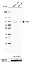
- Western blot analysis in A-431 cells transfected with control siRNA, target specific siRNA probe #1, using Anti-SUN2 antibody. Remaining relative intensity is presented. Loading control: Anti-GAPDH.
- Sample type
- Human
- Protocol
- Protocol

Supportive validation
- Submitted by
- Atlas Antibodies (provider)
- Main image

- Experimental details
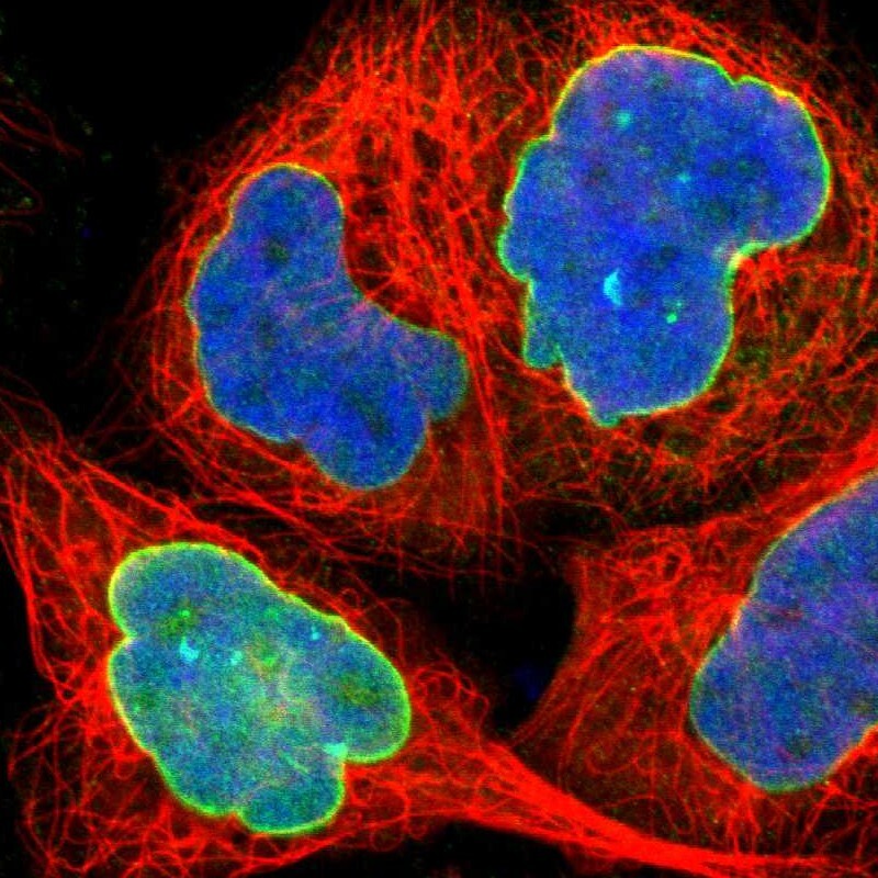
- Immunofluorescent staining of human cell line A-431 shows positivity in nuclear membrane.
- Sample type
- Human