Explore
 Validate
 Learn
Learn Flow cytometry
Antibody data
- Antibody Data
- Antigen structure
- References [1]
- Comments [0]
- Validations
- Flow cytometry [1]
- Other assay [4]
Submit
Validation data
Reference
Comment
Report error
- Product number
- 48-0238-41 - Provider product page

- Provider
- Invitrogen Antibodies
- Product name
- CD23 Monoclonal Antibody (EBVCS2), eFluor™ 450, eBioscience™
- Antibody type
- Monoclonal
- Antigen
- Other
- Description
- Description: The EBVCS2 monoclonal antibody reacts with human CD23, a 45 kDa type II transmembrane glycoprotein. CD23 is expressed on mature B cells, mantle zone B cells, follicular dendritic cells and at low levels on T, NK, langerhans cells and platelets. Expression of CD23 is upregulated upon B cell activation, and soluble forms of the antigen have been reported to be biologically active. CD23 is a low affinity receptor for IgE and is thought to play a role in the regulation of IgE response and B cell activation. CD21 and the alpha subunit of CD11b and CD11c bind to CD23.
- Antibody clone number
- EBVCS2
- Concentration
- 5 µL/Test
Submitted references Interleukin-25 fails to activate STAT6 and induce alternatively activated macrophages.
Stolfi C, Caruso R, Franzè E, Sarra M, De Nitto D, Rizzo A, Pallone F, Monteleone G
Immunology 2011 Jan;132(1):66-77
Immunology 2011 Jan;132(1):66-77
No comments: Submit comment
Supportive validation
- Submitted by
- Invitrogen Antibodies (provider)
- Main image
- Experimental details
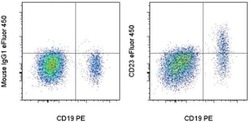
- Staining of normal human peripheral blood cells with Anti-Human CD19 PE (Product # 12-0199-80) and Mouse IgG1 K Isotype Control eFluor® 450 (Product # 48-4714-82) (left) or Anti-Human CD23 eFluor® 450 (right). Cells in the lymphocyte gate were used for analysis.

Supportive validation
- Submitted by
- Invitrogen Antibodies (provider)
- Main image
- Experimental details
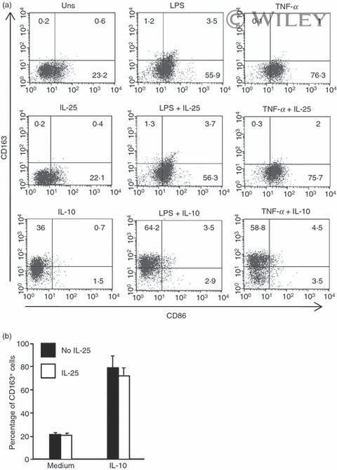
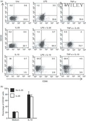
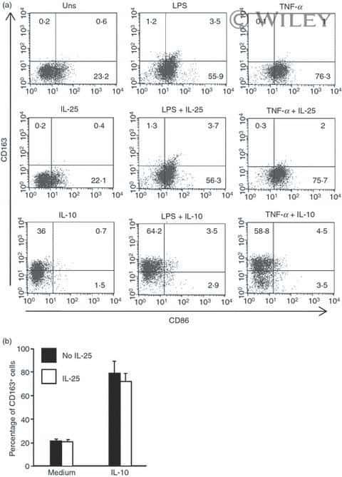
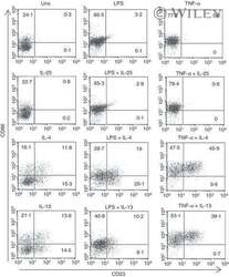
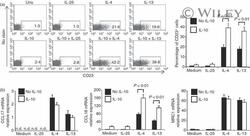
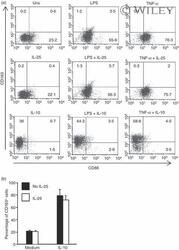
- 5 Interleukin-25 (IL-25) does not alter the expression of CD163 and CD86 in human CD14 + cells. (a) Representative dot-plots showing the expression of CD163 and CD86 in CD14 + cells pre-incubated with medium, IL-25 (50 ng/mL) or IL-10 (25 ng/ml) for 30 min and then either left unstimulated (Uns) or stimulated with lipopolysaccharide (LPS; 100 ng/ml) or tumour necrosis factor-alpha (TNF-alpha; 10 ng/ml). After 12 hr, CD23 and CD163 expression was assessed by flow cytometry. Numbers indicate the percentage of positive cells within the designated quadrants. One of three representative experiments in which similar results were obtained is shown. (b) Induction of CD163 by IL-10 is not influenced by IL-25. Representative histograms showing the percentage of CD14 + cells positive for CD163. Cells were pre-incubated with IL-25 (50 ng/ml) for 6 hr and then stimulated with IL-10 (25 ng/ml) for a further 24 hr. Data indicate the mean +- SD of three experiments.
- Submitted by
- Invitrogen Antibodies (provider)
- Main image
- Experimental details
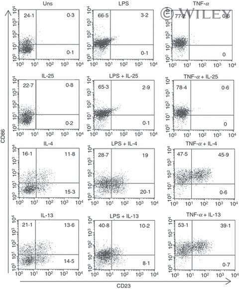
- 2 Interleukin-4 (IL-4) and IL-13, but not IL-25, enhance the fraction of CD23-expressing cells. Representative dot-plots showing the expression of CD23 and CD86 in CD14 + cells pre-incubated with medium, IL-25, IL-4 or IL-13 (50 ng/ml) for 30 min and then stimulated or not with lipopolysaccharide (LPS; 100 ng/ml) or tumour necrosis factor-alpha (TNF-alpha; 10 ng/ml) for a further 12 hr. CD23 and CD86 expression was assessed by flow-cytometry. Numbers indicate the percentage of positive cells within the designated quadrants. One of three representative experiments in which similar results were obtained is shown.
- Submitted by
- Invitrogen Antibodies (provider)
- Main image
- Experimental details
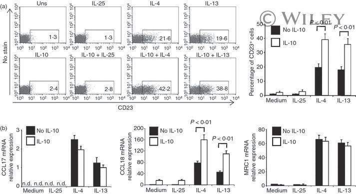
- 4 Interleukin-25 (IL-25) fails to induce alternatively activated macrophage-associated markers in human monocytes pre-incubated with IL-10. (a) Representative dot-plots showing the expression of CD23 in CD14 + cells pre-incubated with medium or IL-10 (25 ng/ml) for 30 min and then either left unstimulated (Uns) or stimulated with IL-25, IL-4 or IL-13 (50 ng/ml). After 12 hr, CD23 expression was assessed by flow-cytometry. Numbers indicate the percentage of positive cells within the designated areas. One of three representative experiments in which similar results were obtained is shown. Right inset. Representative histograms showing the percentage of CD14 + cells expressing CD23 and cultured as indicated above. Data indicate the mean +- SD of three experiments (b) Representative histograms showing CCL17, CCL18 and MRC1 mRNA expression in CD14 + cells, pre-incubated or not with IL-10 for 30 min and then stimulated with IL-25, IL-4 or IL-13 for a further 3 hr. RNA was extracted and amplified by real-time PCR. Levels are normalized to beta-actin and indicate the mean +- SD of three experiments. nd = not detectable.
- Submitted by
- Invitrogen Antibodies (provider)
- Main image
- Experimental details
- 5 Interleukin-25 (IL-25) does not alter the expression of CD163 and CD86 in human CD14 + cells. (a) Representative dot-plots showing the expression of CD163 and CD86 in CD14 + cells pre-incubated with medium, IL-25 (50 ng/mL) or IL-10 (25 ng/ml) for 30 min and then either left unstimulated (Uns) or stimulated with lipopolysaccharide (LPS; 100 ng/ml) or tumour necrosis factor-alpha (TNF-alpha; 10 ng/ml). After 12 hr, CD23 and CD163 expression was assessed by flow cytometry. Numbers indicate the percentage of positive cells within the designated quadrants. One of three representative experiments in which similar results were obtained is shown. (b) Induction of CD163 by IL-10 is not influenced by IL-25. Representative histograms showing the percentage of CD14 + cells positive for CD163. Cells were pre-incubated with IL-25 (50 ng/ml) for 6 hr and then stimulated with IL-10 (25 ng/ml) for a further 24 hr. Data indicate the mean +- SD of three experiments.