 Explore
 Validate
 Learn
Learn Western blot
Antibody data
- Antibody Data
- Antigen structure
- References [0]
- Comments [0]
- Validations
- Western blot [1]
- Immunocytochemistry [1]
- Immunoprecipitation [1]
- Immunohistochemistry [1]
Submit
Validation data
Reference
Comment
Report error
- Product number
- AF6440 - Provider product page

- Provider
- R&D Systems
- Product name
- Rat Endoglin/CD105 Antibody
- Antibody type
- Polyclonal
- Description
- Antigen Affinity-purified. Detects rat Endoglin/CD105 in direct ELISAs and Western blots. In direct ELISAs, approximately 75% cross reactivity with recombinant mouse Endoglin is observed and approximately 50% cross reactivity with recombinant human Endoglin is observed.
- Reactivity
- Rat
- Host
- Goat
- Conjugate
- Unconjugated
- Antigen sequence
Q6Q3E8- Isotype
- IgG
- Vial size
- 100 ug
- Storage
- Use a manual defrost freezer and avoid repeated freeze-thaw cycles. 12 months from date of receipt, -20 to -70 °C as supplied. 1 month, 2 to 8 °C under sterile conditions after reconstitution. 6 months, -20 to -70 °C under sterile conditions after reconstitution.
No comments: Submit comment
Supportive validation
- Submitted by
- R&D Systems (provider)
- Main image

- Experimental details
- Detection of Rat Endoglin/CD105 by Western Blot. Western blot shows lysates of rat kidney tissue. PVDF membrane was probed with 1 µg/mL of Goat Anti-Rat Endoglin/CD105 Antigen Affinity-purified Polyclonal Antibody (Catalog # AF6440) followed by HRP-conjugated Anti-Goat IgG Secondary Antibody (Catalog # HAF017). A specific band was detected for Endoglin/CD105 at approximately 90 kDa (as indicated). This experiment was conducted under reducing conditions and using Immunoblot Buffer Group 1.

Supportive validation
- Submitted by
- R&D Systems (provider)
- Main image

- Experimental details
- Endoglin/CD105 in Rat Mesenchymal Stem Cells. Endoglin/CD105 was detected in immersion fixed undifferentiated rat mesenchymal stem cells using Goat Anti-Rat Endoglin/CD105 Antigen Affinity-purified Polyclonal Antibody (Catalog # AF6440) at 10 µg/mL for 3 hours at room temperature. Cells were stained using the NorthernLights™ 557-conjugated Anti-Goat IgG Secondary Antibody (red; Catalog # NL001) and counterstained with DAPI (blue). Specific staining was localized to the transmembrane. View our protocol for Fluorescent ICC Staining of Stem Cells on Coverslips.

Supportive validation
- Submitted by
- R&D Systems (provider)
- Main image
- Experimental details
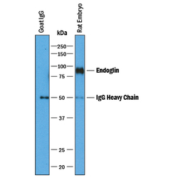
- Immunoprecipitation of Rat Endoglin/CD105. Endoglin/CD105 was immunoprecipitated from 400 µg of rat embryo tissue lysates using 4 µg of Goat Anti-Rat Endoglin/CD105 Antigen Affinity-purified Polyclonal Antibody (Catalog # AF6440) coated on 4 wells of a 96 well plate (Corning Costar EIA/RIA). Rat embryo tissue lysates or control buffer were added to the wells and incubated for 2 hours at room temperature. Immunoprecipitated Endoglin/CD105 was detected by Western blot under reducing conditions using 1 µg/mL Goat Anti-Rat Endoglin/CD105 Antigen Affinity-purified Polyclonal Antibody (Catalog # AF6440) and Immunoblot Buffer Group 1. View our recommended buffer recipes for Immunoprecipitation.

Supportive validation
- Submitted by
- R&D Systems (provider)
- Main image

- Experimental details
- Endoglin/CD105 in Rat Kidney. Endoglin/CD105 was detected in perfusion fixed frozen sections of rat kidney using Goat Anti-Rat Endoglin/CD105 Antigen Affinity-purified Polyclonal Antibody (Catalog # AF6440) at 1 µg/mL overnight at 4 °C. Tissue was stained using the NorthernLights™ 557-conjugated Anti-Goat IgG Secondary Antibody (red; Catalog # NL001) and counterstained with DAPI (blue). Specific staining was localized to endothelial cells in glomeruli. View our protocol for Fluorescent IHC Staining of Frozen Tissue Sections.
