 Explore
 Validate
 Learn
Learn Flow cytometry
Antibody data
- Antibody Data
- Antigen structure
- References [14]
- Comments [0]
- Validations
- Flow cytometry [1]
- Other assay [5]
Submit
Validation data
Reference
Comment
Report error
- Product number
- MHCD10504 - Provider product page

- Provider
- Invitrogen Antibodies
- Product name
- CD105 Monoclonal Antibody (SN6), PE
- Antibody type
- Monoclonal
- Antigen
- Other
- Description
- R-phycoerythrin (PE) is a stable and highly soluble phycobiliprotein which provides maximal absorbance and fluorescence without susceptibility to internal or external fluorescence quenching, thus providing an exceptional quantum yields and molar extinction coefficients.
- Reactivity
- Human
- Host
- Mouse
- Conjugate
- Yellow dye
- Isotype
- IgG
- Antibody clone number
- SN6
- Vial size
- 500 µL
- Storage
- 4° C, store in dark
Submitted references Direct comparison of different therapeutic cell types susceptibility to inflammatory cytokines associated with COVID-19 acute lung injury.
Effects of triptolide on bone marrow-derived mesenchymal stem cells from patients with multiple myeloma.
Endoglin: a novel target for therapeutic intervention in acute leukemias revealed in xenograft mouse models.
Extensive Characterization and Comparison of Endothelial Cells Derived from Dermis and Adipose Tissue: Potential Use in Tissue Engineering.
Long-Term Expansion in Platelet Lysate Increases Growth of Peripheral Blood-Derived Endothelial-Colony Forming Cells and Their Growth Factor-Induced Sprouting Capacity.
The isolation and culture of endothelial colony-forming cells from human and rat lungs.
PDE3A mutations cause autosomal dominant hypertension with brachydactyly.
Induced Pluripotent Stem Cells to Model Human Fibrodysplasia Ossificans Progressiva.
Functionality of endothelial cells and pericytes from human pluripotent stem cells demonstrated in cultured vascular plexus and zebrafish xenografts.
Characterization and functionality of proliferative human Sertoli cells.
Aging and replicative senescence have related effects on human stem and progenitor cells.
Replicative senescence of mesenchymal stem cells: a continuous and organized process.
Mesenchymal stem cell features of Ewing tumors.
Immunophenotype of human adipose-derived cells: temporal changes in stromal-associated and stem cell-associated markers.
Vaka R, Khan S, Ye B, Risha Y, Parent S, Courtman D, Stewart DJ, Davis DR
Stem cell research & therapy 2022 Jan 15;13(1):20
Stem cell research & therapy 2022 Jan 15;13(1):20
Effects of triptolide on bone marrow-derived mesenchymal stem cells from patients with multiple myeloma.
Wu H, Wu Y, Ren L, Zhai W, Jiang Y, Guo S, Tao D, Su C, Chen Z, Jiang H
Experimental and therapeutic medicine 2019 May;17(5):3291-3298
Experimental and therapeutic medicine 2019 May;17(5):3291-3298
Endoglin: a novel target for therapeutic intervention in acute leukemias revealed in xenograft mouse models.
Dourado KMC, Baik J, Oliveira VKP, Beltrame M, Yamamoto A, Theuer CP, Figueiredo CAV, Verneris MR, Perlingeiro RCR
Blood 2017 May 4;129(18):2526-2536
Blood 2017 May 4;129(18):2526-2536
Extensive Characterization and Comparison of Endothelial Cells Derived from Dermis and Adipose Tissue: Potential Use in Tissue Engineering.
Monsuur HN, Weijers EM, Niessen FB, Gefen A, Koolwijk P, Gibbs S, van den Broek LJ
PloS one 2016;11(11):e0167056
PloS one 2016;11(11):e0167056
Long-Term Expansion in Platelet Lysate Increases Growth of Peripheral Blood-Derived Endothelial-Colony Forming Cells and Their Growth Factor-Induced Sprouting Capacity.
Tasev D, van Wijhe MH, Weijers EM, van Hinsbergh VW, Koolwijk P
PloS one 2015;10(6):e0129935
PloS one 2015;10(6):e0129935
The isolation and culture of endothelial colony-forming cells from human and rat lungs.
Alphonse RS, Vadivel A, Zhong S, McConaghy S, Ohls R, Yoder MC, Thébaud B
Nature protocols 2015 Nov;10(11):1697-708
Nature protocols 2015 Nov;10(11):1697-708
PDE3A mutations cause autosomal dominant hypertension with brachydactyly.
Maass PG, Aydin A, Luft FC, Schächterle C, Weise A, Stricker S, Lindschau C, Vaegler M, Qadri F, Toka HR, Schulz H, Krawitz PM, Parkhomchuk D, Hecht J, Hollfinger I, Wefeld-Neuenfeld Y, Bartels-Klein E, Mühl A, Kann M, Schuster H, Chitayat D, Bialer MG, Wienker TF, Ott J, Rittscher K, Liehr T, Jordan J, Plessis G, Tank J, Mai K, Naraghi R, Hodge R, Hopp M, Hattenbach LO, Busjahn A, Rauch A, Vandeput F, Gong M, Rüschendorf F, Hübner N, Haller H, Mundlos S, Bilginturan N, Movsesian MA, Klussmann E, Toka O, Bähring S
Nature genetics 2015 Jun;47(6):647-53
Nature genetics 2015 Jun;47(6):647-53
Induced Pluripotent Stem Cells to Model Human Fibrodysplasia Ossificans Progressiva.
Cai J, Orlova VV, Cai X, Eekhoff EMW, Zhang K, Pei D, Pan G, Mummery CL, Ten Dijke P
Stem cell reports 2015 Dec 8;5(6):963-970
Stem cell reports 2015 Dec 8;5(6):963-970
Functionality of endothelial cells and pericytes from human pluripotent stem cells demonstrated in cultured vascular plexus and zebrafish xenografts.
Orlova VV, Drabsch Y, Freund C, Petrus-Reurer S, van den Hil FE, Muenthaisong S, Dijke PT, Mummery CL
Arteriosclerosis, thrombosis, and vascular biology 2014 Jan;34(1):177-86
Arteriosclerosis, thrombosis, and vascular biology 2014 Jan;34(1):177-86
Characterization and functionality of proliferative human Sertoli cells.
Chui K, Trivedi A, Cheng CY, Cherbavaz DB, Dazin PF, Huynh AL, Mitchell JB, Rabinovich GA, Noble-Haeusslein LJ, John CM
Cell transplantation 2011;20(5):619-35
Cell transplantation 2011;20(5):619-35
Aging and replicative senescence have related effects on human stem and progenitor cells.
Wagner W, Bork S, Horn P, Krunic D, Walenda T, Diehlmann A, Benes V, Blake J, Huber FX, Eckstein V, Boukamp P, Ho AD
PloS one 2009 Jun 9;4(6):e5846
PloS one 2009 Jun 9;4(6):e5846
Replicative senescence of mesenchymal stem cells: a continuous and organized process.
Wagner W, Horn P, Castoldi M, Diehlmann A, Bork S, Saffrich R, Benes V, Blake J, Pfister S, Eckstein V, Ho AD
PloS one 2008 May 21;3(5):e2213
PloS one 2008 May 21;3(5):e2213
Mesenchymal stem cell features of Ewing tumors.
Tirode F, Laud-Duval K, Prieur A, Delorme B, Charbord P, Delattre O
Cancer cell 2007 May;11(5):421-9
Cancer cell 2007 May;11(5):421-9
Immunophenotype of human adipose-derived cells: temporal changes in stromal-associated and stem cell-associated markers.
Mitchell JB, McIntosh K, Zvonic S, Garrett S, Floyd ZE, Kloster A, Di Halvorsen Y, Storms RW, Goh B, Kilroy G, Wu X, Gimble JM
Stem cells (Dayton, Ohio) 2006 Feb;24(2):376-85
Stem cells (Dayton, Ohio) 2006 Feb;24(2):376-85
No comments: Submit comment
Supportive validation
- Submitted by
- Invitrogen Antibodies (provider)
- Main image

- Experimental details
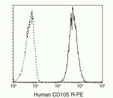
- Antigen Specificity: According to the literature this antibody recognizes the CD105 antigen. Also known as endoglin, this antigen is expressed on endothelial cells, some bone marrow cells and activated macrophages.
- Conjugate
- Yellow dye
Supportive validation
- Submitted by
- Invitrogen Antibodies (provider)
- Main image
- Experimental details
- NULL
- Conjugate
- Yellow dye

- Submitted by
- Invitrogen Antibodies (provider)
- Main image

- Experimental details
- NULL
- Conjugate
- Yellow dye

- Submitted by
- Invitrogen Antibodies (provider)
- Main image

- Experimental details
- NULL
- Conjugate
- Yellow dye

- Submitted by
- Invitrogen Antibodies (provider)
- Main image
- Experimental details
- NULL
- Conjugate
- Yellow dye

- Submitted by
- Invitrogen Antibodies (provider)
- Main image

- Experimental details
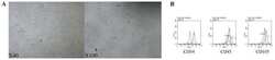
- Fig 2 Phenotypical characterization of PB-ECFCs. PB-ECFCs monolayers expanded in EGM-2 medium supplemented with 10%PL were assessed for the presence of endothelial cell markers by immunofluorescence cytochemistry ( A-C ) and flow cytometry as well as for uptake of Dil-Ac-LDL ( D ). For immunofluorescence cytochemistry staining, cells were seeded on glass cover slips, fixed and stained with antibody against CD31, VE-cadherin or vWF. Cell nuclei were visualized with DAPI staining. Cells were positive for CD31 ( A , green), VE-cadherin ( B , green), and vWF ( C , green). Cell nuclei appear blue. ( D ): Incorporation of Dil-Ac-LDL by PB-ECFCs (red spots, cell nuclei stained blue with DAPI). Panel E :Flow cytometry characterization of PB-ECFCs for CD31, CD34, CD309, CD144, CD146, CD105, CD14, CD45, and CD133. Plots depict control isotype IgG staining (black histograms) versus specific antibody staining (empty histograms).
- Conjugate
- Yellow dye
