 Explore
 Validate
 Learn
Learn Flow cytometry
Antibody data
- Antibody Data
- Antigen structure
- References [5]
- Comments [0]
- Validations
- Flow cytometry [1]
- Other assay [4]
Submit
Validation data
Reference
Comment
Report error
- Product number
- MHCD10520 - Provider product page

- Provider
- Invitrogen Antibodies
- Product name
- CD105 Monoclonal Antibody (SN6), Alexa Fluor™ 488
- Antibody type
- Monoclonal
- Antigen
- Other
- Description
- Based on our testing, publications, and results reported from customers, the Alexa Fluor® 488 dye provides the best fluorescein (FITC) substitute.
- Reactivity
- Human
- Host
- Mouse
- Conjugate
- Green dye
- Isotype
- IgG
- Antibody clone number
- SN6
- Vial size
- 500 µL
- Storage
- 4° C, store in dark
Submitted references Novel low shear 3D bioreactor for high purity mesenchymal stem cell production.
The effects of 3D culture on the expansion and maintenance of nucleus pulposus progenitor cell multipotency.
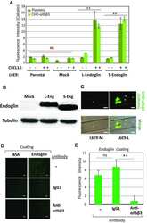
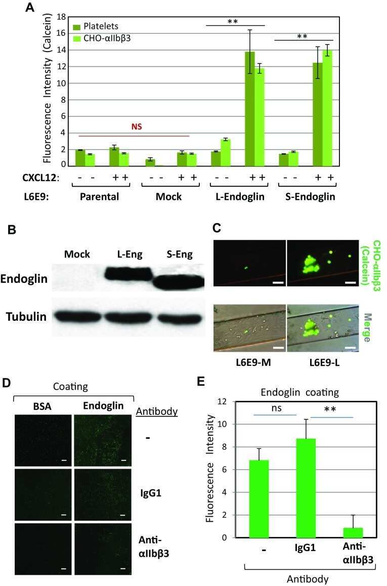
Human endoglin as a potential new partner involved in platelet-endothelium interactions.
Myogenic potential of human alveolar mucosa derived cells.
Octacalcium phosphate ceramics combined with gingiva-derived stromal cells for engineered functional bone grafts.
Burns AB, Doris C, Vehar K, Saxena V, Bardliving C, Shamlou PA, Phillips MI
PloS one 2021;16(6):e0252575
PloS one 2021;16(6):e0252575
The effects of 3D culture on the expansion and maintenance of nucleus pulposus progenitor cell multipotency.
Guerrero J, Häckel S, Croft AS, Albers CE, Gantenbein B
JOR spine 2021 Mar;4(1):e1131
JOR spine 2021 Mar;4(1):e1131
Human endoglin as a potential new partner involved in platelet-endothelium interactions.
Rossi E, Pericacho M, Bachelot-Loza C, Pidard D, Gaussem P, Poirault-Chassac S, Blanco FJ, Langa C, González-Manchón C, Novoa JML, Smadja DM, Bernabeu C
Cellular and molecular life sciences : CMLS 2018 Apr;75(7):1269-1284
Cellular and molecular life sciences : CMLS 2018 Apr;75(7):1269-1284
Myogenic potential of human alveolar mucosa derived cells.
Zorin VL, Pulin AA, Eremin II, Korsakov IN, Zorina AI, Khromova NV, Sokova OI, Kotenko KV, Kopnin PB
Cell cycle (Georgetown, Tex.) 2017 Mar 19;16(6):545-555
Cell cycle (Georgetown, Tex.) 2017 Mar 19;16(6):545-555
Octacalcium phosphate ceramics combined with gingiva-derived stromal cells for engineered functional bone grafts.
Zorin VL, Komlev VS, Zorina AI, Khromova NV, Solovieva EV, Fedotov AY, Eremin II, Kopnin PB
Biomedical materials (Bristol, England) 2014 Aug 28;9(5):055005
Biomedical materials (Bristol, England) 2014 Aug 28;9(5):055005
No comments: Submit comment
Supportive validation
- Submitted by
- Invitrogen Antibodies (provider)
- Main image
- Experimental details
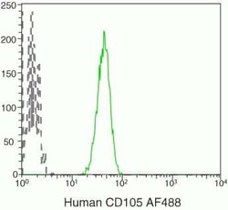
- U937 cells were stained with Alexa Fluor® 488-conjugated anti-human CD105 monoclonal antibody (Product # MHCD10520). The negative profile represents unstained cells.
- Conjugate
- Green dye

Supportive validation
- Submitted by
- Invitrogen Antibodies (provider)
- Main image
- Experimental details
- NULL
- Conjugate
- Green dye
- Submitted by
- Invitrogen Antibodies (provider)
- Main image
- Experimental details
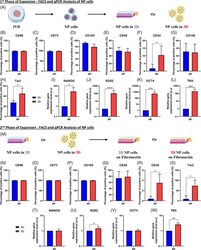
- FIGURE 2 Analysis of NP cells after the first and second phase of expansion. A, Schematic representation of the first phase of expansion of nucleus pulposus (NP) cells. Briefly, after dissection and digestion of the NP tissue from the intervertebral disk (IVD), NP cells were seeded in two-dimensional (2D) or three-dimensional (3D) into alginate beads. B-H, Quantification by flow cytometry analysis of the amount of NP cells positive for different markers after culture in 2D or 3D for 1 week of culture. B-H, Percentage of NP cells positive in 2D or 3D for, B, CD90 marker, C, CD73 marker, D, CD105 marker, E, CD45 marker, F, CD34 marker, G, CD146 marker, and, H, Tie2 marker. I-L, Quantification of the relative gene on NP cells for different genes after culture in 2D or 3D for 1 week of culture. I, Relative gene expression of NANOG in NP cells in 2D or 3D. J, Relative gene expression of SOX2 in NP cells in 2D or 3D. K, Relative gene expression of OCT4 in NP cells in 2D or 3D. L, Relative gene expression of TEK in NP cells in 2D or 3D. M, Schematic representation of the second phase of expansion of NP cells. Briefly, after the first phase of expansion, NP cells cultured in 2D or 3D were putting back in the fibronectin-coated flask/surface. N-S, Quantification by flow cytometry analysis of the amount of NP cells (previously cultivated in 2D or 3D) positive for different markers after culture in the fibronectin-coated surface for 1 week of culture for, N, CD90 marker, O, CD73 marker,
- Conjugate
- Green dye

- Submitted by
- Invitrogen Antibodies (provider)
- Main image
- Experimental details
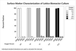
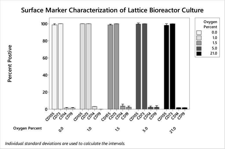
- 10.1371/journal.pone.0252575.g007 Fig 7 Flow cytometry of hMSC from bioreactors harvested day seven from varying oxygen tension. CD105, CD73, CD19, and CD14 stained cells were analyzed using flow cytometry. No significant differences were found in biomarker expression after preconditioning using 0.0%, 1.0%, 1.5%. 5.0%, and 21% gasses.
- Conjugate
- Green dye
- Submitted by
- Invitrogen Antibodies (provider)
- Main image

- Experimental details
- 10.1371/journal.pone.0252575.g008 Fig 8 hMSC biomarker characterization using flow cytometry. Cells were cultured in both static and dynamic bioreactor conditions, and compared using CD105, C73, CD19, and CD14 staining. B) Overlaid flow cytometry image of 1.5% O 2 primed hMSC cultures from day seven dynamic bioreactors showing positive markers and negative markers.
- Conjugate
- Green dye
