 Explore
 Validate
 Learn
Learn Western blot
 Immunocytochemistry
Antibody data
- Antibody Data
- Antigen structure
- References [1]
- Comments [0]
- Validations
- Western blot [1]
- Immunohistochemistry [1]
- Flow cytometry [2]
Submit
Validation data
Reference
Comment
Report error
- Product number
- NB100-77666 - Provider product page

- Provider
- Novus Biologicals
- Proper citation
- Novus Cat#NB100-77666, RRID:AB_1083158
- Product name
- Rat Monoclonal Endoglin/CD105 Antibody
- Antibody type
- Monoclonal
- Description
- Protein G purified.
- Reactivity
- Mouse
- Host
- Rat
- Antigen sequence
Endothelial cell selective TGB1 bin
ding molecule-endoglin- Isotype
- IgG
- Vial size
- 0.5 mg
- Concentration
- 1.0 mg/ml
- Storage
- Store at 4C short term. Aliquot and store at -20C long term. Avoid freeze-thaw cycles.
Submitted references Targeting angiogenesis for radioimmunotherapy with a 177Lu-labeled antibody.
Ehlerding EB, Lacognata S, Jiang D, Ferreira CA, Goel S, Hernandez R, Jeffery JJ, Theuer CP, Cai W
European journal of nuclear medicine and molecular imaging 2018 Jan;45(1):123-131
European journal of nuclear medicine and molecular imaging 2018 Jan;45(1):123-131
No comments: Submit comment
Supportive validation
- Submitted by
- Novus Biologicals (provider)
- Main image
- Experimental details
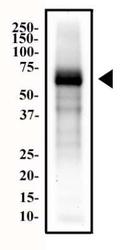
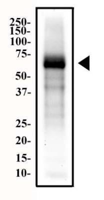
- Western Blot: Endoglin/CD105 Antibody (MJ7/18) [NB100-77666] - Total protein from mouse placenta was separated on a 12% gel by SDS-PAGE, transferred to PVDF membrane and blocking in 5% non-fat milk in TBST. The membrane was probed with 2 ug/ml anti-CD105 (NB100-77666) in 1% milk/TBST and detected with an anti-rat HRP conjugated secondary antibody using chemiluminescence.
Supportive validation
- Submitted by
- Novus Biologicals (provider)
- Main image
- Experimental details
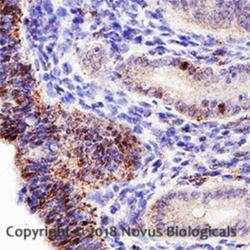
- Immunohistochemistry-Paraffin: Endoglin/CD105 Antibody (MJ7/18) [NB100-77666] - Analysis of a FFPE mouse ductus deference using a 1:50 dilution of CD105 antibody (clone MJ7/18) on a Bond Rx autostainer (Leica Biosystems). The assay involved 20 minutes of heat induced antigen retrieval (HIER) using 10mM sodium citrate buffer (pH 6.0) and endogenous peroxidase quenching with peroxide block. The sections were incubated with primary antibody for 30 minutes and Bond Polymer Refine Detection (Leica Biosystems) with DAB was used for signal development followed by counterstaining with hematoxylin. Whole slide scanning and capturing of representative images (20X) was performed using Aperio AT2 (Leica Biosystems). Staining was observed in the pseudostratified columnar epithelial cells. Staining was performed by Histowiz.

Supportive validation
- Submitted by
- Novus Biologicals (provider)
- Main image

- Experimental details
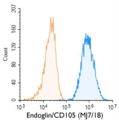
- Flow (Cell Surface): Endoglin/CD105 Antibody (MJ7/18) [NB100-77666] - A surface stain was performed on MS-1 Cells with Endoglin/CD105 (MJ7/18) antibody NB100-77666 (blue) and a matched isotype control (orange). Cells were incubated in an antibody dilution of 1 ug/mL for 20 minutes at room temperature, followed by rat F(ab)2 IgG (H+L) APC-conjugated secondary antibody (F0113, R&D Systems).
- Submitted by
- Novus Biologicals (provider)
- Main image
- Experimental details
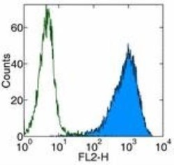
- Flow Cytometry: Endoglin/CD105 Antibody (MJ7/18) [NB100-77666] - Analysis of Biotin conjugate of NB100-77666. Surface staining of bEND.3 cell line with 0.25 ug of Anti-Mouse CD105 (Endoglin) Biotin followed by Streptavidin PE. Appropriate isotype controls were used (open histogram). Total viable cells were used for analysis.
