Explore
 Validate
 Learn
Learn Western blot
 ELISA
ELISAAntibody data
- Antibody Data
- Antigen structure
- References [1]
- Comments [0]
- Validations
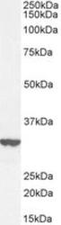
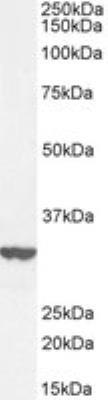
- Western blot [1]
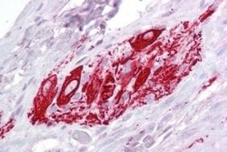
- Immunohistochemistry [1]
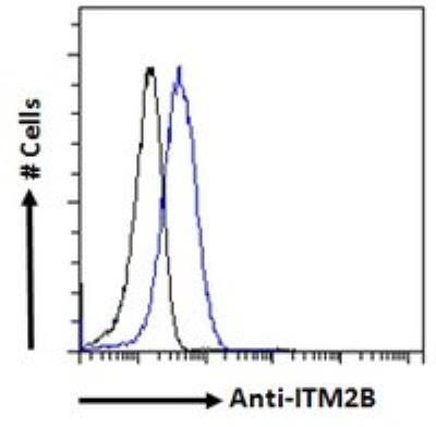
- Flow cytometry [1]
Submit
Validation data
Reference
Comment
Report error
- Product number
- NBP1-36963 - Provider product page

- Provider
- Novus Biologicals
- Proper citation
- Novus Cat#NBP1-36963, RRID:AB_2129810
- Product name
- Goat Polyclonal ITM2B Antibody
- Antibody type
- Polyclonal
- Description
- Immunogen affinity purified.
- Reactivity
- Human
- Host
- Goat
- Antigen sequence
internal region- Isotype
- IgG
- Vial size
- 0.1 mg
- Concentration
- 0.5 mg/ml
- Storage
- Store at -20C. Avoid freeze-thaw cycles.
Submitted references Understanding the development of human bladder cancer by using a whole-organ genomic mapping strategy.
Majewski T, Lee S, Jeong J, Yoon DS, Kram A, Kim MS, Tuziak T, Bondaruk J, Lee S, Park WS, Tang KS, Chung W, Shen L, Ahmed SS, Johnston DA, Grossman HB, Dinney CP, Zhou JH, Harris RA, Snyder C, Filipek S, Narod SA, Watson P, Lynch HT, Gazdar A, Bar-Eli M, Wu XF, McConkey DJ, Baggerly K, Issa JP, Benedict WF, Scherer SE, Czerniak B
Laboratory investigation; a journal of technical methods and pathology 2008 Jul;88(7):694-721
Laboratory investigation; a journal of technical methods and pathology 2008 Jul;88(7):694-721
No comments: Submit comment
Supportive validation
- Submitted by
- Novus Biologicals (provider)
- Main image
- Experimental details
- Western Blot: ITM2B Antibody [NBP1-36963] - HepG2 cell lysate (35 ug protein in RIPA buffer). Antibody at 0.3 ug/mL. Detected by chemiluminescence.
Supportive validation
- Submitted by
- Novus Biologicals (provider)
- Main image
- Experimental details
- Immunohistochemistry-Paraffin: ITM2B Antibody [NBP1-36963] - Human small intestine tissue. Antibody at 2.5 ug/mL. Steamed antigen retrieval with citrate buffer pH 6, AP-staining.

Supportive validation
- Submitted by
- Novus Biologicals (provider)
- Main image
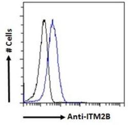
- Experimental details
- Flow Cytometry: ITM2B Antibody [NBP1-36963] - Paraformaldehyde fixed HeLa cells (blue line), permeabilized with 0.5% Triton. Primary antibody at 10 ug/mL, 1 hr incubation followed by Alexa Fluor 488 secondary antibody (1 ug/mL). IgG control: Unimmunized goat IgG (black line) followed by Alexa Fluor 488 secondary antibody.