Explore
 Validate
 Learn
Learn Western blot
Antibody data
- Antibody Data
- Antigen structure
- References [11]
- Comments [0]
- Validations
- Western blot [14]
- Immunocytochemistry [1]
- Immunohistochemistry [4]
Submit
Validation data
Reference
Comment
Report error
- Product number
- GTX104763 - Provider product page

- Provider
- GeneTex
- Proper citation
- GeneTex Cat#GTX104763, RRID:AB_1240586
- Product name
- PD-L1 antibody
- Antibody type
- Polyclonal
- Reactivity
- Human
- Host
- Rabbit
Submitted references Nuclear localization of PD-L1: artifact or reality?
Overexpressed miR-195 attenuated immune escape of diffuse large B-cell lymphoma by targeting PD-L1.
Eradication of Triple-Negative Breast Cancer Cells by Targeting Glycosylated PD-L1.
Metformin Promotes Antitumor Immunity via Endoplasmic-Reticulum-Associated Degradation of PD-L1.
Soluble PD-L1 with PD-1-binding capacity exists in the plasma of patients with non-small cell lung cancer.
Tumor-infiltrating lymphocyte composition, organization and PD-1/ PD-L1 expression are linked in breast cancer.
A combination of anti-PD-L1 mAb plus Lm-LLO-E6 vaccine efficiently suppresses tumor growth and metastasis in HPV-infected cancers.
An increase in BAG-1 by PD-L1 confers resistance to tyrosine kinase inhibitor in non-small cell lung cancer via persistent activation of ERK signalling.
Intestine-Specific Homeobox Gene ISX Integrates IL6 Signaling, Tryptophan Catabolism, and Immune Suppression.
Upregulation of PD-L1 and APE1 is associated with tumorigenesis and poor prognosis of gastric cancer.
High PD-L1 Expression Correlates with Metastasis and Poor Prognosis in Oral Squamous Cell Carcinoma.
Polioudaki H, Chantziou A, Kalyvianaki K, Malamos P, Notas G, Mavroudis D, Kampa M, Castanas E, Theodoropoulos PA
Cellular oncology (Dordrecht) 2019 Apr;42(2):237-242
Cellular oncology (Dordrecht) 2019 Apr;42(2):237-242
Overexpressed miR-195 attenuated immune escape of diffuse large B-cell lymphoma by targeting PD-L1.
He B, Yan F, Wu C
Biomedicine & pharmacotherapy = Biomedecine & pharmacotherapie 2018 Feb;98:95-101
Biomedicine & pharmacotherapy = Biomedecine & pharmacotherapie 2018 Feb;98:95-101
Eradication of Triple-Negative Breast Cancer Cells by Targeting Glycosylated PD-L1.
Li CW, Lim SO, Chung EM, Kim YS, Park AH, Yao J, Cha JH, Xia W, Chan LC, Kim T, Chang SS, Lee HH, Chou CK, Liu YL, Yeh HC, Perillo EP, Dunn AK, Kuo CW, Khoo KH, Hsu JL, Wu Y, Hsu JM, Yamaguchi H, Huang TH, Sahin AA, Hortobagyi GN, Yoo SS, Hung MC
Cancer cell 2018 Feb 12;33(2):187-201.e10
Cancer cell 2018 Feb 12;33(2):187-201.e10
Metformin Promotes Antitumor Immunity via Endoplasmic-Reticulum-Associated Degradation of PD-L1.
Cha JH, Yang WH, Xia W, Wei Y, Chan LC, Lim SO, Li CW, Kim T, Chang SS, Lee HH, Hsu JL, Wang HL, Kuo CW, Chang WC, Hadad S, Purdie CA, McCoy AM, Cai S, Tu Y, Litton JK, Mittendorf EA, Moulder SL, Symmans WF, Thompson AM, Piwnica-Worms H, Chen CH, Khoo KH, Hung MC
Molecular cell 2018 Aug 16;71(4):606-620.e7
Molecular cell 2018 Aug 16;71(4):606-620.e7
Soluble PD-L1 with PD-1-binding capacity exists in the plasma of patients with non-small cell lung cancer.
Takeuchi M, Doi T, Obayashi K, Hirai A, Yoneda K, Tanaka F, Iwai Y
Immunology letters 2018 Apr;196:155-160
Immunology letters 2018 Apr;196:155-160
Tumor-infiltrating lymphocyte composition, organization and PD-1/ PD-L1 expression are linked in breast cancer.
Buisseret L, Garaud S, de Wind A, Van den Eynden G, Boisson A, Solinas C, Gu-Trantien C, Naveaux C, Lodewyckx JN, Duvillier H, Craciun L, Veys I, Larsimont D, Piccart-Gebhart M, Stagg J, Sotiriou C, Willard-Gallo K
Oncoimmunology 2017;6(1):e1257452
Oncoimmunology 2017;6(1):e1257452
A combination of anti-PD-L1 mAb plus Lm-LLO-E6 vaccine efficiently suppresses tumor growth and metastasis in HPV-infected cancers.
Lin PL, Cheng YM, Wu DW, Huang YJ, Lin HC, Chen CY, Lee H
Cancer medicine 2017 Sep;6(9):2052-2062
Cancer medicine 2017 Sep;6(9):2052-2062
An increase in BAG-1 by PD-L1 confers resistance to tyrosine kinase inhibitor in non-small cell lung cancer via persistent activation of ERK signalling.
Lin PL, Wu TC, Wu DW, Wang L, Chen CY, Lee H
European journal of cancer (Oxford, England : 1990) 2017 Nov;85:95-105
European journal of cancer (Oxford, England : 1990) 2017 Nov;85:95-105
Intestine-Specific Homeobox Gene ISX Integrates IL6 Signaling, Tryptophan Catabolism, and Immune Suppression.
Wang LT, Chiou SS, Chai CY, Hsi E, Yokoyama KK, Wang SN, Huang SK, Hsu SH
Cancer research 2017 Aug 1;77(15):4065-4077
Cancer research 2017 Aug 1;77(15):4065-4077
Upregulation of PD-L1 and APE1 is associated with tumorigenesis and poor prognosis of gastric cancer.
Qing Y, Li Q, Ren T, Xia W, Peng Y, Liu GL, Luo H, Yang YX, Dai XY, Zhou SF, Wang D
Drug design, development and therapy 2015;9:901-9
Drug design, development and therapy 2015;9:901-9
High PD-L1 Expression Correlates with Metastasis and Poor Prognosis in Oral Squamous Cell Carcinoma.
Lin YM, Sung WW, Hsieh MJ, Tsai SC, Lai HW, Yang SM, Shen KH, Chen MK, Lee H, Yeh KT, Chen CJ
PloS one 2015;10(11):e0142656
PloS one 2015;10(11):e0142656
No comments: Submit comment
Supportive validation
- Submitted by
- GeneTex (provider)
- Main image

- Experimental details
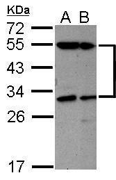
- Sample (30 ug of whole cell lysate) A: K562 B: THP-1 12% SDS PAGE GTX100562 diluted at 1:1000
- Validation comment
- WB
- Submitted by
- GeneTex (provider)
- Main image
- Experimental details
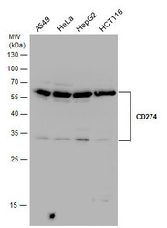
- CD274 antibody detects CD274 protein by western blot analysis. Various whole cell extracts (30 ?g) were separated by 12% SDS-PAGE, and the membrane was blotted with CD274 antibody (GTX104763) diluted by 1:500.
- Validation comment
- WB
- Submitted by
- GeneTex (provider)
- Main image
- Experimental details
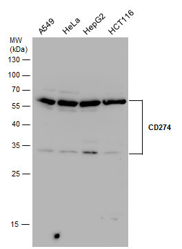
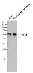
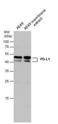
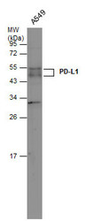
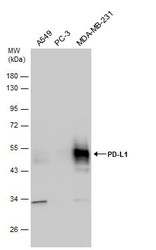
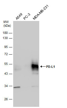
- PD-L1 antibody detects PD-L1 protein by western blot analysis. A549 whole cell extracts and membrane extracts (30 ?g) were separated by 12% SDS-PAGE, and the membrane was blotted with PD-L1 antibody (GTX104763) diluted at 1:500.
- Validation comment
- WB
- Submitted by
- GeneTex (provider)
- Main image
- Experimental details
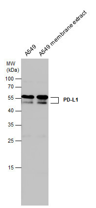
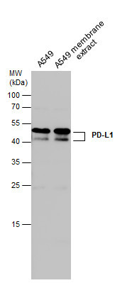
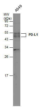
- A549 whole cell and membrane extracts (30 ?g) were separated by 12% SDS-PAGE, and the membrane was blotted with PD-L1 antibody (GTX104763) diluted at 1:500. The HRP-conjugated anti-rabbit IgG antibody (GTX213110-01) was used to detect the primary antibody.
- Submitted by
- GeneTex (provider)
- Main image
- Experimental details
- Whole cell extract (30 ?g) was separated by 12% SDS-PAGE, and the membrane was blotted with PD-L1 antibody (GTX104763) diluted at 1:500. The HRP-conjugated anti-rabbit IgG antibody (GTX213110-01) was used to detect the primary antibody.
- Submitted by
- GeneTex (provider)
- Main image
- Experimental details
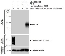
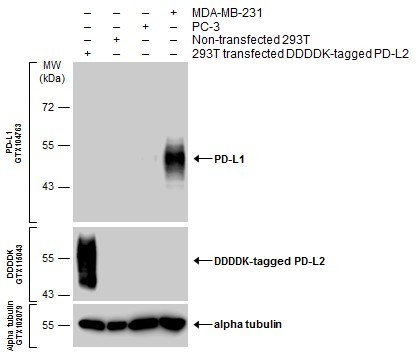
- Various whole cell extracts were separated by 10% SDS-PAGE, and the membranes were blotted with PD-L1 antibody (GTX104763) diluted at 1:600 and with DDDDK tag antibody (GTX115043) diluted at 1:3000 to detect DDDDK-tagged PD-L2. The HRP-conjugated anti-rabbit IgG antibody (GTX213110-01) was used to detect the primary antibody.
- Submitted by
- GeneTex (provider)
- Main image
- Experimental details
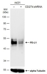
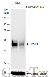
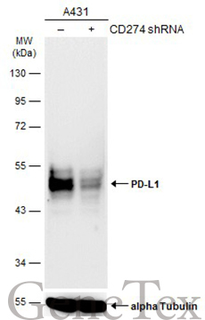
- Non-transfected (¡V) and transfected (+) A431 whole cell extracts (30 ?g) were separated by 10% SDS-PAGE, and the membrane was blotted with PD-L1 antibody (GTX104763) diluted at 1:1000. The HRP-conjugated anti-rabbit IgG antibody (GTX213110-01) was used to detect the primary antibody.
- Submitted by
- GeneTex (provider)
- Main image
- Experimental details
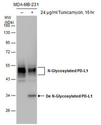
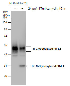
- Untreated (¡V) and treated (+) MDA-MB-231 whole cell extracts (30 ?g) were separated by 10% SDS-PAGE, and the membrane was blotted with PD-L1 antibody (GTX104763) diluted at 1:1000.
- Submitted by
- GeneTex (provider)
- Main image
- Experimental details
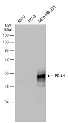
- Various whole cell extracts (30 ?g) were separated by 12% SDS-PAGE, and the membrane was blotted with PD-L1 antibody (GTX104763) diluted at 1:2000. The HRP-conjugated anti-rabbit IgG antibody (GTX213110-01) was used to detect the primary antibody.
- Submitted by
- GeneTex (provider)
- Main image
- Experimental details
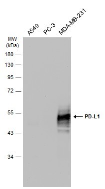
- Various whole cell extracts (30 ?g) were separated by 10% SDS-PAGE, and the membrane was blotted with PD-L1 antibody (GTX104763) diluted at 1:2000. The HRP-conjugated anti-rabbit IgG antibody (GTX213110-01) was used to detect the primary antibody.
- Submitted by
- GeneTex (provider)
- Main image
- Experimental details
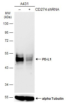
- Non-transfected (¡V) and transfected (+) A431 whole cell extracts (30 ?g) were separated by 10% SDS-PAGE, and the membrane was blotted with PD-L1 antibody (GTX104763) diluted at 1:1000. The HRP-conjugated anti-rabbit IgG antibody (GTX213110-01) was used to detect the primary antibody.
- Submitted by
- GeneTex (provider)
- Main image
- Experimental details
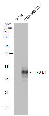
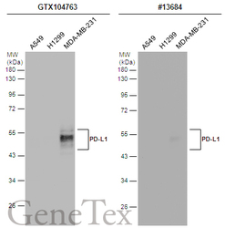
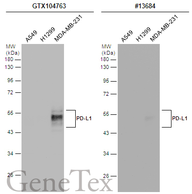
- Various whole cell extracts (30 ?g) were separated by 12% SDS-PAGE, and the membranes were blotted with PD-L1 antibody (GTX104763) diluted at 1:2000 and competitor's antibody (CST#13684) diluted by 1:500. The HRP-conjugated anti-rabbit IgG antibody (GTX213110-01) was used to detect the primary antibody.
- Submitted by
- GeneTex (provider)
- Main image
- Experimental details
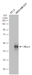
- Various whole cell extracts (30 ?g) were separated by 10% SDS-PAGE, and the membrane was blotted with PD-L1 antibody (GTX104763) diluted at 1:2000.
- Submitted by
- GeneTex (provider)
- Main image
- Experimental details
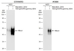
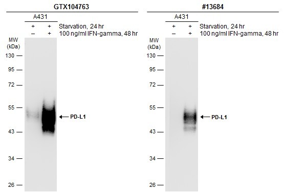
- Untreated (¡V) and treated (+) A431 whole cell extracts (30 ?g) were separated by 10% SDS-PAGE, and the membranes were blotted with PD-L1 antibody (GTX104763) diluted at 1:1200 and competitor's antibody (CST#13684) diluted at 1:500. The HRP-conjugated anti-rabbit IgG antibody (GTX213110-01) was used to detect the primary antibody.
Supportive validation
- Submitted by
- GeneTex (provider)
- Main image
- Experimental details
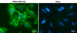
- PD-L1 antibody detects PD-L1 protein at cell membrane in MDA-MB-231 cells by immunofluorescent analysis.Sample: MDA-MB-231 (left) and HeLa (right) cells.Green: PD-L1 protein stained by PD-L1 antibody (GTX104763) diluted at 1:1000.Blue: Hoechst 33342 staining.Scale bar = 10 £gm.

Supportive validation
- Submitted by
- GeneTex (provider)
- Main image

- Experimental details
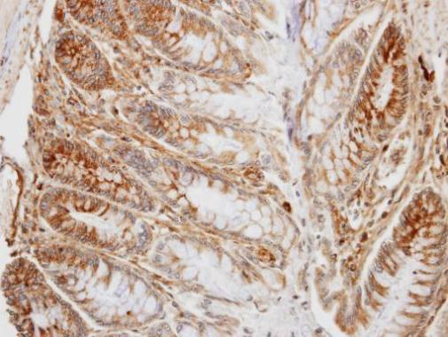
- Immunohistochemical analysis of paraffin-embedded human colon, using PD-L1(GTX104763) antibody at 1:100 dilution.
- Submitted by
- GeneTex (provider)
- Main image
- Experimental details
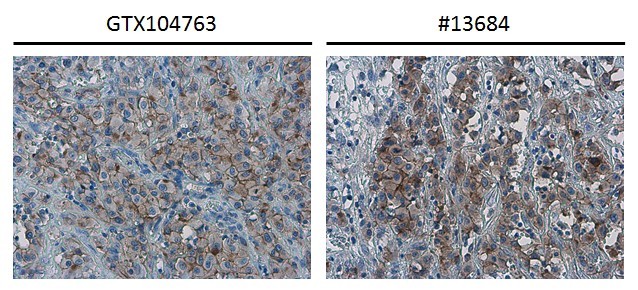
- PD-L1 antibody detects PD-L1 protein at cell membrane in human ovarian carcinoma by immunohistochemical analysis. Antibodies: PD-L1 antibody (GTX104763) diluted at 1:1000, and competitor's antibody diluted at 1:50.
- Submitted by
- GeneTex (provider)
- Main image
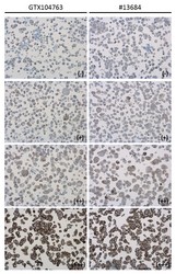
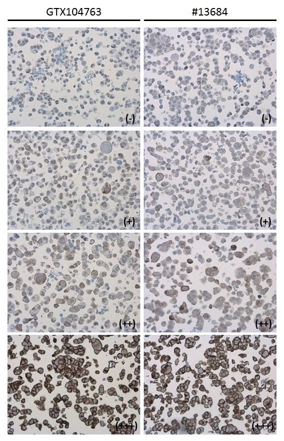
- Experimental details
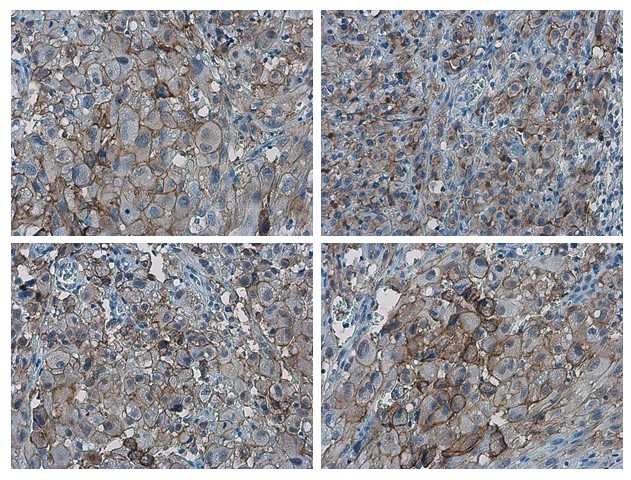
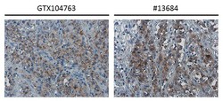
- PD-L1 antibody detects PD-L1 protein at cell membrane in PD-L1 protein-expressing cell lines by immunohistochemical analysis. Antibodies: PD-L1 antibody (GTX104763) diluted at 1:1000, and competitor's antibody diluted at 1:50. Samples: Negative (-), low positive (+), intermediate positive (++) and strong positive (+++) cell line cores assessed using Quantitative Digital Pathology.
- Submitted by
- GeneTex (provider)
- Main image
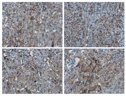
- Experimental details
- PD-L1 antibody detects PD-L1 proteinat cell membrane in human ovarian carcinoma by immunohistochemical analysis. Sample: Paraffin-embedded human ovarian carcinoma. PD-L1 antibody (GTX104763) diluted at 1:1000.