 Explore
 Validate
 Learn
Learn Western blot
 Immunohistochemistry
Antibody data
- Antibody Data
- Antigen structure
- References [14]
- Comments [0]
- Validations
- Western blot [1]
- Flow cytometry [2]
Submit
Validation data
Reference
Comment
Report error
- Product number
- AF1019 - Provider product page

- Provider
- R&D Systems
- Product name
- Mouse PD-L1 Antibody
- Antibody type
- Polyclonal
- Description
- Antigen Affinity-purified. Detects mouse PD-L1/B7-H1 in direct ELISAs and Western blots. In direct ELISAs, less than 20% cross-reactivity with recombinant human PD-L1/B7-H1 is observed.
- Reactivity
- Mouse
- Host
- Goat
- Conjugate
- Unconjugated
- Antigen sequence
Q9EP73- Isotype
- IgG
- Vial size
- 100 ug
- Concentration
- LYOPH
- Storage
- Use a manual defrost freezer and avoid repeated freeze-thaw cycles. 12 months from date of receipt, -20 to -70 °C as supplied. 1 month, 2 to 8 °C under sterile conditions after reconstitution. 6 months, -20 to -70 °C under sterile conditions after reconstitution.
Submitted references IL-17C-mediated innate inflammation decreases the response to PD-1 blockade in a model of Kras-driven lung cancer.
Interferon-gamma drives programmed death-ligand 1 expression on islet β cells to limit T cell function during autoimmune diabetes.
PD-L1 checkpoint inhibition and anti-CTLA-4 whole tumor cell vaccination counter adaptive immune resistance: A mouse neuroblastoma model that mimics human disease.
Tumor-Localized Secretion of Soluble PD1 Enhances Oncolytic Virotherapy.
PD-1/PD-L1 Blockade Enhances T-cell Activity and Antitumor Efficacy of Imatinib in Gastrointestinal Stromal Tumors.
Hormonal vitamin D up-regulates tissue-specific PD-L1 and PD-L2 surface glycoprotein expression in humans but not mice.
Dependence of Glomerulonephritis Induction on Novel Intraglomerular Alternatively Activated Bone Marrow-Derived Macrophages and Mac-1 and PD-L1 in Lupus-Prone NZM2328 Mice.
Cell autonomous or systemic EGFR blockade alters the immune-environment in squamous cell carcinomas.
β-Cell-targeted blockage of PD1 and CTLA4 pathways prevents development of autoimmune diabetes and acute allogeneic islets rejection.
IFN-γ from lymphocytes induces PD-L1 expression and promotes progression of ovarian cancer.
Endogenous retinoids in the pathogenesis of alopecia areata.
Anti-programmed cell death 1 antibody reduces CD4+PD-1+ T cells and relieves the lupus-like nephritis of NZB/W F1 mice.
Altered availability of PD-1/PD ligands is associated with the failure to control autoimmunity in NOD mice.
Establishment of NOD-Pdcd1-/- mice as an efficient animal model of type I diabetes.
Ritzmann F, Jungnickel C, Vella G, Kamyschnikow A, Herr C, Li D, Menger MM, Angenendt A, Hoth M, Lis A, Bals R, Beisswenger C
Scientific reports 2019 Jul 17;9(1):10353
Scientific reports 2019 Jul 17;9(1):10353
Interferon-gamma drives programmed death-ligand 1 expression on islet β cells to limit T cell function during autoimmune diabetes.
Osum KC, Burrack AL, Martinov T, Sahli NL, Mitchell JS, Tucker CG, Pauken KE, Papas K, Appakalai B, Spanier JA, Fife BT
Scientific reports 2018 May 29;8(1):8295
Scientific reports 2018 May 29;8(1):8295
PD-L1 checkpoint inhibition and anti-CTLA-4 whole tumor cell vaccination counter adaptive immune resistance: A mouse neuroblastoma model that mimics human disease.
Srinivasan P, Wu X, Basu M, Rossi C, Sandler AD
PLoS medicine 2018 Jan;15(1):e1002497
PLoS medicine 2018 Jan;15(1):e1002497
Tumor-Localized Secretion of Soluble PD1 Enhances Oncolytic Virotherapy.
Bartee MY, Dunlap KM, Bartee E
Cancer research 2017 Jun 1;77(11):2952-2963
Cancer research 2017 Jun 1;77(11):2952-2963
PD-1/PD-L1 Blockade Enhances T-cell Activity and Antitumor Efficacy of Imatinib in Gastrointestinal Stromal Tumors.
Seifert AM, Zeng S, Zhang JQ, Kim TS, Cohen NA, Beckman MJ, Medina BD, Maltbaek JH, Loo JK, Crawley MH, Rossi F, Besmer P, Antonescu CR, DeMatteo RP
Clinical cancer research : an official journal of the American Association for Cancer Research 2017 Jan 15;23(2):454-465
Clinical cancer research : an official journal of the American Association for Cancer Research 2017 Jan 15;23(2):454-465
Hormonal vitamin D up-regulates tissue-specific PD-L1 and PD-L2 surface glycoprotein expression in humans but not mice.
Dimitrov V, Bouttier M, Boukhaled G, Salehi-Tabar R, Avramescu RG, Memari B, Hasaj B, Lukacs GL, Krawczyk CM, White JH
The Journal of biological chemistry 2017 Dec 15;292(50):20657-20668
The Journal of biological chemistry 2017 Dec 15;292(50):20657-20668
Dependence of Glomerulonephritis Induction on Novel Intraglomerular Alternatively Activated Bone Marrow-Derived Macrophages and Mac-1 and PD-L1 in Lupus-Prone NZM2328 Mice.
Sung SJ, Ge Y, Dai C, Wang H, Fu SM, Sharma R, Hahn YS, Yu J, Le TH, Okusa MD, Bolton WK, Lawler JR
Journal of immunology (Baltimore, Md. : 1950) 2017 Apr 1;198(7):2589-2601
Journal of immunology (Baltimore, Md. : 1950) 2017 Apr 1;198(7):2589-2601
Cell autonomous or systemic EGFR blockade alters the immune-environment in squamous cell carcinomas.
Mascia F, Schloemann DT, Cataisson C, McKinnon KM, Krymskaya L, Wolcott KM, Yuspa SH
International journal of cancer 2016 Dec 1;139(11):2593-7
International journal of cancer 2016 Dec 1;139(11):2593-7
β-Cell-targeted blockage of PD1 and CTLA4 pathways prevents development of autoimmune diabetes and acute allogeneic islets rejection.
El Khatib MM, Sakuma T, Tonne JM, Mohamed MS, Holditch SJ, Lu B, Kudva YC, Ikeda Y
Gene therapy 2015 May;22(5):430-8
Gene therapy 2015 May;22(5):430-8
IFN-γ from lymphocytes induces PD-L1 expression and promotes progression of ovarian cancer.
Abiko K, Matsumura N, Hamanishi J, Horikawa N, Murakami R, Yamaguchi K, Yoshioka Y, Baba T, Konishi I, Mandai M
British journal of cancer 2015 Apr 28;112(9):1501-9
British journal of cancer 2015 Apr 28;112(9):1501-9
Endogenous retinoids in the pathogenesis of alopecia areata.
Duncan FJ, Silva KA, Johnson CJ, King BL, Szatkiewicz JP, Kamdar SP, Ong DE, Napoli JL, Wang J, King LE Jr, Whiting DA, McElwee KJ, Sundberg JP, Everts HB
The Journal of investigative dermatology 2013 Feb;133(2):334-43
The Journal of investigative dermatology 2013 Feb;133(2):334-43
Anti-programmed cell death 1 antibody reduces CD4+PD-1+ T cells and relieves the lupus-like nephritis of NZB/W F1 mice.
Kasagi S, Kawano S, Okazaki T, Honjo T, Morinobu A, Hatachi S, Shimatani K, Tanaka Y, Minato N, Kumagai S
Journal of immunology (Baltimore, Md. : 1950) 2010 Mar 1;184(5):2337-47
Journal of immunology (Baltimore, Md. : 1950) 2010 Mar 1;184(5):2337-47
Altered availability of PD-1/PD ligands is associated with the failure to control autoimmunity in NOD mice.
Yadav D, Hill N, Yagita H, Azuma M, Sarvetnick N
Cellular immunology 2009;258(2):161-71
Cellular immunology 2009;258(2):161-71
Establishment of NOD-Pdcd1-/- mice as an efficient animal model of type I diabetes.
Wang J, Yoshida T, Nakaki F, Hiai H, Okazaki T, Honjo T
Proceedings of the National Academy of Sciences of the United States of America 2005 Aug 16;102(33):11823-8
Proceedings of the National Academy of Sciences of the United States of America 2005 Aug 16;102(33):11823-8
No comments: Submit comment
Supportive validation
- Submitted by
- R&D Systems (provider)
- Main image

- Experimental details
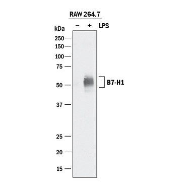
- Detection of Mouse PD-L1/B7-H1 by Western Blot. Western blot shows lysates of RAW 264.7 mouse monocyte/macrophage cell line untreated (-) or treated (+) with 10 μg/mL LPS for 4 hours. PVDF membrane was probed with 0.5 µg/mL of Goat Anti-Mouse PD-L1/B7-H1 Antigen Affinity-purified Polyclonal Antibody (Catalog # AF1019) followed by HRP-conjugated Anti-Goat IgG Secondary Antibody (Catalog # HAF017). A specific band was detected for PD-L1/B7-H1 at approximately 50-55 kDa (as indicated). This experiment was conducted under reducing conditions and using Immunoblot Buffer Group 1.
Supportive validation
- Submitted by
- R&D Systems (provider)
- Main image

- Experimental details
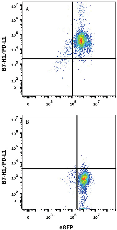
- Detection of PD-L1/B7-H1 in HEK293 Human Cell Line Transfected with Mouse PD-L1/B7-H1 and eGFP by Flow Cytometry. HEK293 human embryonic kidney cell line transfected with either (A) mouse PD-L1/B7-H1 or (B) irrelevant transfectants and eGFP was stained with Goat Anti-Mouse PD-L1/B7-H1 Antigen Affinity-purified Polyclonal Antibody (Catalog # AF1019) followed by Allophycocyanin-conjugated Anti-Goat IgG Secondary Antibody (Catalog # F0108). Quadrant markers were set based on control antibody staining (Catalog # AB-108-C). View our protocol for Staining Membrane-associated Proteins.
- Submitted by
- R&D Systems (provider)
- Main image
- Experimental details
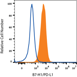
- Detection of PD-L1/B7-H1 in RAW 264.7 Mouse Cell Line by Flow Cytometry. RAW 264.7 mouse monocyte/macrophage cell line either treated with LPS overnight (filled histogram) or untreated (open histogram) was stained with Goat Anti-Mouse PD-L1/B7-H1 Antigen Affinity-purified Polyclonal Antibody (Catalog # AF1019), followed by Allophycocyanin-conjugated Anti-Goat IgG Secondary Antibody (Catalog # F0108). View our protocol for Staining Membrane-associated Proteins.
