Explore
 Validate
 Learn
Learn Western blot
Antibody data
- Antibody Data
- Antigen structure
- References [2]
- Comments [0]
- Validations
- Western blot [1]
- Immunohistochemistry [1]
- Flow cytometry [2]
Submit
Validation data
Reference
Comment
Report error
- Product number
- MAB90781-100 - Provider product page

- Provider
- R&D Systems
- Product name
- Mouse PD-L1/B7-H1 Antibody
- Antibody type
- Monoclonal
- Description
- Protein A or G purified from cell culture supernatant. Detects mouse PD-L1/B7-H1 in direct ELISAs and Western blots.
- Reactivity
- Mouse
- Host
- Rabbit
- Conjugate
- Unconjugated
- Antigen sequence
Q9EP73- Isotype
- IgG
- Antibody clone number
- 2096C
- Vial size
- 100 ug
- Storage
- Use a manual defrost freezer and avoid repeated freeze-thaw cycles. 12 months from date of receipt, -20 to -70 °C as supplied. 1 month, 2 to 8 °C under sterile conditions after reconstitution. 6 months, -20 to -70 °C under sterile conditions after reconstitution.
Submitted references Human Biofield Therapy and the Growth of Mouse Lung Carcinoma.
The Circadian Clock Controls Immune Checkpoint Pathway in Sepsis.
Yang P, Jiang Y, Rhea PR, Coway T, Chen D, Gagea M, Harribance SL, Cohen L
Integrative cancer therapies 2019 Jan-Dec;18:1534735419840797
Integrative cancer therapies 2019 Jan-Dec;18:1534735419840797
The Circadian Clock Controls Immune Checkpoint Pathway in Sepsis.
Deng W, Zhu S, Zeng L, Liu J, Kang R, Yang M, Cao L, Wang H, Billiar TR, Jiang J, Xie M, Tang D
Cell reports 2018 Jul 10;24(2):366-378
Cell reports 2018 Jul 10;24(2):366-378
No comments: Submit comment
Supportive validation
- Submitted by
- R&D Systems (provider)
- Main image

- Experimental details
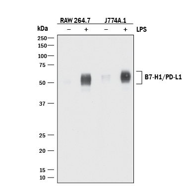
- Detection of Mouse PD-L1/B7-H1 by Western Blot. Western blot shows lysates of RAW 264.7 mouse monocyte/macrophage cell line and J774A.1 mouse reticulum cell sarcoma macrophage cell line untreated (-) or treated (+) with 10 μg/mL LPS for 4 hours. PVDF membrane was probed with 2 µg/mL of Rabbit Anti-Mouse PD-L1/B7-H1 Monoclonal Antibody (Catalog # MAB90781) followed by HRP-conjugated Anti-Rabbit IgG Secondary Antibody (Catalog # HAF008). A specific band was detected for PD-L1/B7-H1 at approximately 50-55 kDa (as indicated). This experiment was conducted under reducing conditions and using Immunoblot Buffer Group 1.
Supportive validation
- Submitted by
- R&D Systems (provider)
- Main image
- Experimental details
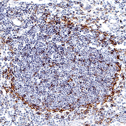
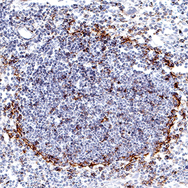
- PD-L1/B7-H1 in Mouse Thymus. PD-L1/B7-H1 was detected in perfusion fixed frozen sections of mouse thymus using Rabbit Anti-Mouse PD-L1/B7-H1 Monoclonal Antibody (Catalog # MAB90781) at 5 µg/mL for 1 hour at room temperature followed by incubation with the Anti-Rabbit IgG VisUCyte™ HRP Polymer Antibody (Catalog # VC003). Tissue was stained using DAB (brown) and counterstained with hematoxylin (blue). Specific staining was localized to thymocytes. View our protocol for IHC Staining with VisUCyte HRP Polymer Detection Reagents.
Supportive validation
- Submitted by
- R&D Systems (provider)
- Main image

- Experimental details
- Detection of PD-L1/B7-H1 in HEK293 Human Cell Line Transfected with Mouse PD-L1/B7-H1 and eGFP by Flow Cytometry. HEK293 human embryonic kidney cell line transfected with either (A) mouse PD-L1/B7-H1 or (B) irrelevant transfectants and eGFP was stained with Rabbit Anti-Mouse PD-L1/B7-H1 Monoclonal Antibody (Catalog # MAB90781) followed by Allophycocyanin-conjugated Anti-Rabbit IgG Secondary Antibody (Catalog # F0111). Quadrant markers were set based on control antibody staining (Catalog # MAB1050). View our protocol for Staining Membrane-associated Proteins.

- Submitted by
- R&D Systems (provider)
- Main image
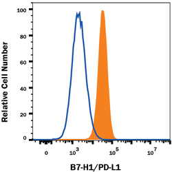
- Experimental details
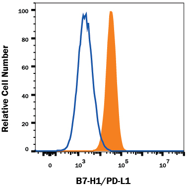
- Detection of PD-L1/B7-H1 in RAW 264.7 Mouse Cell Line by Flow Cytometry. RAW 264.7 mouse monocyte/macrophage cell line either treated with LPS overnight (filled histogram) or untreated (open histogram) was stained with Rabbit Anti-Mouse PD-L1/B7-H1 Monoclonal Antibody (Catalog # MAB90781), followed by Allophycocyanin-conjugated Anti-Rabbit IgG Secondary Antibody (Catalog # F0111). View our protocol for Staining Membrane-associated Proteins.