Explore
 Validate
 Learn
Learn Western blot
Antibody data
- Antibody Data
- Antigen structure
- References [0]
- Comments [0]
- Validations
- Western blot [2]
- Immunohistochemistry [1]
- Flow cytometry [2]
Submit
Validation data
Reference
Comment
Report error
- Product number
- 10-7599-25 - Provider product page

- Provider
- ABEOMICS Inc.
- Product name
- Anti-Human PD-L1 Antibody
- Antibody type
- Monoclonal
- Description
- PD-L1 (CD274/B7-H1) is a critical membrane-bound costimulatory molecule belongs to the B7 superfamily that inhibits immune responses through its receptor, PD-1 and PD-L1 play a key role in the pathogenesis of inflammatory diseases (programmed death 1). It is widely expressed in the mononuclear phagocyte system (MPS), may co-stimulate T cells and regulates inflammatory responses. PD-L1 exerts inflammation regulatory functions via a negative co-stimulatory effect on T cell functions to inhibit cytokine secretion, facilitate apoptosis of activated T cells and induce T cell anergy. Aberrant expression and dysregulation of CD274 have been reported during bacterial infection, inflammation and in numerous autoimmune diseases.
- Reactivity
- Human
- Host
- Mouse
- Conjugate
- Unconjugated
- Antigen sequence
A partial length recombinant protei
n of PD-L1 (amino acid 13-224) was
used as the immunogen for this anti
body.- Isotype
- IgG
- Antibody clone number
- ABM5F25
- Vial size
- 100 µg
- Concentration
- 0.5 mg/ml
- Storage
- Store the antibody at 4°C, stable for 6 months. For long-term storage, store at -20°C. Avoid repeat freez thawing
No comments: Submit comment
Supportive validation
- Submitted by
- ABEOMICS Inc. (provider)
- Main image
- Experimental details
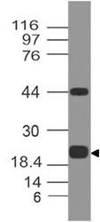
- Western blot analysis of PDL1. Anti-PD-L1 antibody (Clone: ABM5F25) was tested at 0.5 µg/ml on Recombinant lysate.
- Protocol
- Protocol

- Submitted by
- ABEOMICS Inc. (provider)
- Main image
- Experimental details
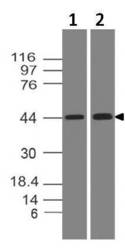
- Western blot analysis of PDL1. Anti-PD-L1 antibody (Clone: ABM5F25) was tested at 2 µg/ml on (1) Daudi and (2) HepG2 lysates.
- Protocol
- Protocol

Supportive validation
- Submitted by
- ABEOMICS Inc. (provider)
- Main image
- Experimental details
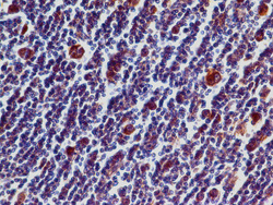
- Immunohistochemical analysis of PD-L1 in Hodkin's Lymphoma tissue using PD-L1 antibody (Clone: ABM5F25) at 5 µg/ml.
- Protocol
- Protocol

Supportive validation
- Submitted by
- ABEOMICS Inc. (provider)
- Main image

- Experimental details
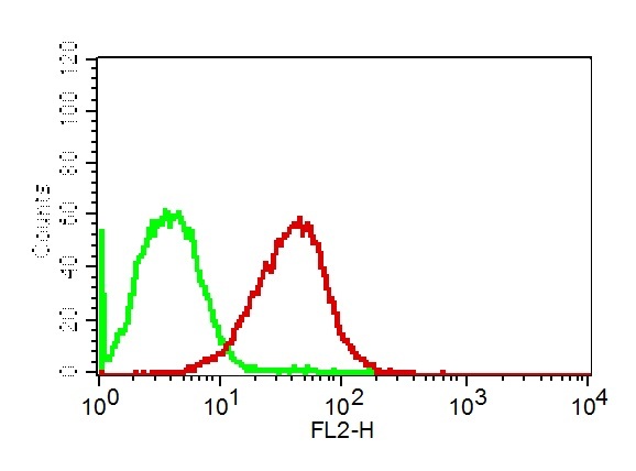
- Cell Surface flow analysis of PD-L1 in 3 day-PHA treated human PBMC cells using 1 µg/10^6 cells of PD-L1 antibody (Clone: ABM5F25). Green represents isotype control; red represents anti-PD-L1 antibody. Goat anti-mouse PE conjugate was used as secondary antibody.
- Protocol
- Protocol

- Submitted by
- ABEOMICS Inc. (provider)
- Main image

- Experimental details
- Cell surface flow analysis of PD-L1 in CHO-PD-L1 transfected cell line using 0.5 µg/10^6 cells of PD-L1 antibody (Clone: ABM5F25). Green represents isotype control; red represents anti-PD-L1 antibody. Goat anti-mouse PE conjugate was used as secondary antibody.
- Protocol
- Protocol