 Explore
 Validate
 Learn
Learn Western blot
 Blocking/Neutralizing
Antibody data
- Antibody Data
- Antigen structure
- References [0]
- Comments [0]
- Validations
- Western blot [1]
- Flow cytometry [1]
Submit
Validation data
Reference
Comment
Report error
- Product number
- MAB1562-100 - Provider product page

- Provider
- R&D Systems
- Product name
- Human PD-L1/B7-H1 Antibody
- Antibody type
- Monoclonal
- Description
- Protein A or G purified from cell culture supernatant. Detects human PD-L1/B7-H1 in direct ELISAs.
- Reactivity
- Human
- Host
- Rabbit
- Conjugate
- Unconjugated
- Antigen sequence
Q9NZQ7- Isotype
- IgG
- Antibody clone number
- 2340D
- Vial size
- 100 ug
- Storage
- Use a manual defrost freezer and avoid repeated freeze-thaw cycles. 12 months from date of receipt, -20 to -70 °C as supplied. 1 month, 2 to 8 °C under sterile conditions after reconstitution. 6 months, -20 to -70 °C under sterile conditions after reconstitution.
No comments: Submit comment
Supportive validation
- Submitted by
- R&D Systems (provider)
- Main image
- Experimental details
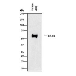
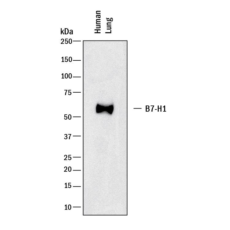
- Detection of human PD-L1/B7-H1 by Western Blot. Western blot shows lysates of human lung tissue. PVDF membrane was probed with 2 µg/mL of Rabbit Anti-Human PD-L1/B7-H1 Monoclonal Antibody (Catalog # MAB1562) followed by HRP-conjugated Anti-Rabbit IgG Secondary Antibody (Catalog # HAF008). A specific band was detected for PD-L1/B7-H1 at approximately 50 kDa (as indicated). This experiment was conducted under reducing conditions and using Immunoblot Buffer Group 1.
Supportive validation
- Submitted by
- R&D Systems (provider)
- Main image
- Experimental details
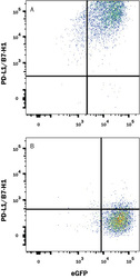
- Detection of PD-L1/B7-H1 in HEK293 Human Cell Line Transfected with Human PD-L1/B7-H1 and eGFP by Flow Cytometry. HEK293 human embryonic kidney cell line transfected with either (A) human PD-L1/B7-H1 or (B) irrelevant protein and eGFP was stained with Rabbit Anti-Human PD-L1/B7-H1 Monoclonal Antibody (Catalog # MAB1562) followed by APC-conjugated Goat anti-Rabbit IgG Secondary Antibody (Catalog # F0111). Quadrant markers were set based on Rabbit IgG control antibody staining (Catalog # MAB1050). View our protocol for Staining Membrane-associated Proteins.
