Explore
 Validate
 Learn
Learn Western blot
 Blocking/Neutralizing
Antibody data
- Antibody Data
- Antigen structure
- References [0]
- Comments [0]
- Validations
- Western blot [1]
- Immunocytochemistry [2]
- Immunohistochemistry [1]
Submit
Validation data
Reference
Comment
Report error
- Product number
- PA5-46985 - Provider product page

- Provider
- Invitrogen Antibodies
- Product name
- LDLR Polyclonal Antibody
- Antibody type
- Polyclonal
- Antigen
- Recombinant full-length protein
- Description
- In direct ELISAs, approximately 15% cross-reactivity with recombinant mouse LDL R is observed. Reconstitute at 0.2 mg/mL in sterile PBS.
- Reactivity
- Human
- Host
- Goat
- Isotype
- IgG
- Vial size
- 100 µg
- Concentration
- 0.2 mg/mL
- Storage
- -20° C, Avoid Freeze/Thaw Cycles
No comments: Submit comment
Supportive validation
- Submitted by
- Invitrogen Antibodies (provider)
- Main image
- Experimental details
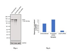
- Knockdown of LDLR was achieved by transfecting PC-3 with LDLR specific siRNAs (Silencer® select Product # s6 and s224007). Western blot analysis (Fig. a) was performed using Whole cell extracts from the LDLR knockdown cells (lane 3), non-targeting scrambled siRNA transfected cells (lane 2) and untransfected cells (lane 1). The blot was probed with LDLR Polyclonal Antibody (Product # PA5-46985, 0.1 µg/ml ) and Rabbit anti-Goat IgG (H+L) Superclonal™ Recombinant Secondary Antibody, HRP (Product # A27014, 1:4000). Densitometric analysis of this western blot is shown in histogram (Fig. b). Decrease in signal upon siRNA mediated knock down confirms that antibody is specific to LDLR.

Supportive validation
- Submitted by
- Invitrogen Antibodies (provider)
- Main image

- Experimental details
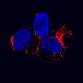
- Immunocytochemistry analysis of LDLR in immersion fixed HepG2 human hepatocellular carcinoma cell line. Samples were incubated in LDLR polyclonal antibody (Product # PA5-46985) using a dilution of 1.7 µg/mL for 3 hours at room temperature followed by NorthernLights™ 557-conjugated Anti-Goat IgG Secondary Antibody (red) and counterstained with DAPI (blue). Specific staining was localized to cytoplasm.
- Submitted by
- Invitrogen Antibodies (provider)
- Main image

- Experimental details
- Immunocytochemistry analysis of LDLR in immersion fixed HepG2 human hepatocellular carcinoma cell line. Samples were incubated in LDLR polyclonal antibody (Product # PA5-46985) using a dilution of 1.7 µg/mL for 3 hours at room temperature followed by NorthernLights™ 557-conjugated Anti-Goat IgG Secondary Antibody (red) and counterstained with DAPI (blue). Specific staining was localized to cytoplasm.

Supportive validation
- Submitted by
- Invitrogen Antibodies (provider)
- Main image
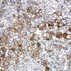
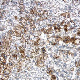
- Experimental details
- Immunohistochemical analysis of LDLR in formalin fixed paraffin-embedded sections of human liver. Samples were incubated in LDLR polyclonal antibody (Product # PA5-46985) using a dilution of 15 µg/mL overnight at 4 °C. Tissue was stained using the Anti-Goat HRP-DAB Cell & Tissue Staining Kit (brown) and counterstained with hematoxylin (blue).