Explore
 Validate
 Learn
Learn Western blot
 Immunocytochemistry
Antibody data
- Antibody Data
- Antigen structure
- References [2]
- Comments [0]
- Validations
- Immunocytochemistry [1]
Submit
Validation data
Reference
Comment
Report error
- Product number
- HPA023026 - Provider product page

- Provider
- Atlas Antibodies
- Proper citation
- Atlas Antibodies Cat#HPA023026, RRID:AB_10963931
- Product name
- Anti-WRAP53
- Antibody type
- Polyclonal
- Description
- Polyclonal Antibody against Human WRAP53, Gene description: WD repeat containing, antisense to TP53, Alternative Gene Names: FLJ10385, TCAB1, WDR79, Validated applications: WB, IHC, ICC, Uniprot ID: Q9BUR4, Storage: Store at +4°C for short term storage. Long time storage is recommended at -20°C.
- Reactivity
- Human
- Host
- Rabbit
- Conjugate
- Unconjugated
- Isotype
- IgG
- Vial size
- 100 µl
- Concentration
- 0.1 mg/ml
- Storage
- Store at +4°C for short term storage. Long time storage is recommended at -20°C.
- Handling
- The antibody solution should be gently mixed before use.
Submitted references Downregulation of the cancer susceptibility protein WRAP53β in epithelial ovarian cancer leads to defective DNA repair and poor clinical outcome
Downregulation of the cancer susceptibility protein WRAP53β in epithelial ovarian cancer leads to defective DNA repair and poor clinical outcome.
Hedström E, Pederiva C, Farnebo J, Nodin B, Jirström K, Brennan D, Farnebo M
Cell Death and Disease 2015 October;6(10)
Cell Death and Disease 2015 October;6(10)
Downregulation of the cancer susceptibility protein WRAP53β in epithelial ovarian cancer leads to defective DNA repair and poor clinical outcome.
Hedström E, Pederiva C, Farnebo J, Nodin B, Jirström K, Brennan DJ, Farnebo M
Cell death & disease 2015 Oct 1;6(10):e1892
Cell death & disease 2015 Oct 1;6(10):e1892
No comments: Submit comment
Supportive validation
- Submitted by
- Atlas Antibodies (provider)
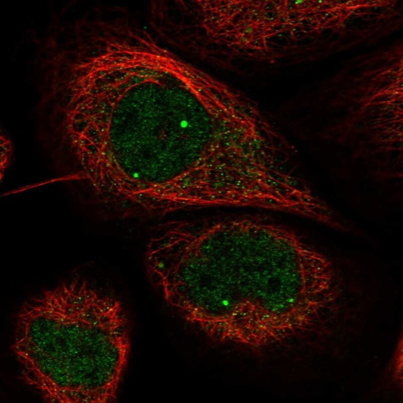
- Main image
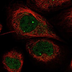
- Experimental details
- Immunofluorescent staining of human cell line A-431 shows localization to nuclear bodies.
- Sample type
- Human