Explore
 Validate
 Learn
LearnNB100-68252
antibody from Novus Biologicals
Targeting: WRAP53
FLJ10385, TCAB1, WDR79
 Western blot
 Immunocytochemistry Immunoprecipitation Immunohistochemistry Chromatin Immunoprecipitation
Antibody data
- Antibody Data
- Antigen structure
- References [8]
- Comments [0]
- Validations
- Western blot [4]
- Immunoprecipitation [1]
- Immunohistochemistry [4]
Submit
Validation data
Reference
Comment
Report error
- Product number
- NB100-68252 - Provider product page

- Provider
- Novus Biologicals
- Proper citation
- Novus Cat#NB100-68252, RRID:AB_1111348
- Product name
- Rabbit Polyclonal WDR79 Antibody
- Antibody type
- Polyclonal
- Description
- Immunogen affinity purified.
- Reactivity
- Human
- Host
- Rabbit
- Isotype
- IgG
- Vial size
- 0.1 ml
- Concentration
- 0.2 mg/ml
- Storage
- Store at 4C. Do not freeze.
Submitted references HnRNP F/H associate with hTERC and telomerase holoenzyme to modulate telomerase function and promote cell proliferation.
N-terminal residues of human dyskerin are required for interactions with telomerase RNA that prevent RNA degradation.
Minimized human telomerase maintains telomeres and resolves endogenous roles of H/ACA proteins, TCAB1, and Cajal bodies.
Human regulator of telomere elongation helicase 1 (RTEL1) is required for the nuclear and cytoplasmic trafficking of pre-U2 RNA.
Dynamics of Human Telomerase Holoenzyme Assembly and Subunit Exchange across the Cell Cycle.
TCAB1: a potential target for diagnosis and therapy of head and neck carcinomas.
Telomerase recruitment requires both TCAB1 and Cajal bodies independently.
WRAP53 promotes cancer cell survival and is a potential target for cancer therapy.
Xu C, Xie N, Su Y, Sun Z, Liang Y, Zhang N, Liu D, Jia S, Xing X, Han L, Li G, Tong T, Chen J
Cell death and differentiation 2020 Jun;27(6):1998-2013
Cell death and differentiation 2020 Jun;27(6):1998-2013
N-terminal residues of human dyskerin are required for interactions with telomerase RNA that prevent RNA degradation.
MacNeil DE, Lambert-Lanteigne P, Autexier C
Nucleic acids research 2019 Jun 4;47(10):5368-5380
Nucleic acids research 2019 Jun 4;47(10):5368-5380
Minimized human telomerase maintains telomeres and resolves endogenous roles of H/ACA proteins, TCAB1, and Cajal bodies.
Vogan JM, Zhang X, Youmans DT, Regalado SG, Johnson JZ, Hockemeyer D, Collins K
eLife 2016 Aug 15;5
eLife 2016 Aug 15;5
Human regulator of telomere elongation helicase 1 (RTEL1) is required for the nuclear and cytoplasmic trafficking of pre-U2 RNA.
Schertzer M, Jouravleva K, Perderiset M, Dingli F, Loew D, Le Guen T, Bardoni B, de Villartay JP, Revy P, Londoño-Vallejo A
Nucleic acids research 2015 Feb 18;43(3):1834-47
Nucleic acids research 2015 Feb 18;43(3):1834-47
Dynamics of Human Telomerase Holoenzyme Assembly and Subunit Exchange across the Cell Cycle.
Vogan JM, Collins K
The Journal of biological chemistry 2015 Aug 28;290(35):21320-35
The Journal of biological chemistry 2015 Aug 28;290(35):21320-35
TCAB1: a potential target for diagnosis and therapy of head and neck carcinomas.
Sun CK, Luo XB, Gou YP, Hu L, Wang K, Li C, Xiang ZT, Zhang P, Kong XL, Zhang CL, Yang Q, Li J, Xiao LY, Li Y, Chen QM
Molecular cancer 2014 Jul 28;13:180
Molecular cancer 2014 Jul 28;13:180
Telomerase recruitment requires both TCAB1 and Cajal bodies independently.
Stern JL, Zyner KG, Pickett HA, Cohen SB, Bryan TM
Molecular and cellular biology 2012 Jul;32(13):2384-95
Molecular and cellular biology 2012 Jul;32(13):2384-95
WRAP53 promotes cancer cell survival and is a potential target for cancer therapy.
Mahmoudi S, Henriksson S, Farnebo L, Roberg K, Farnebo M
Cell death & disease 2011 Jan 13;2:e114
Cell death & disease 2011 Jan 13;2:e114
No comments: Submit comment
Supportive validation
- Submitted by
- Novus Biologicals (provider)
- Main image

- Experimental details
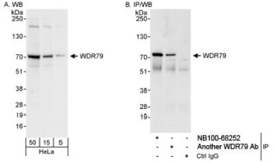
- Western Blot: WDR79 Antibody [NB100-68252] - Whole cell lysate (5, 15 and 50 mcg for WB; 1 mg for IP, 20% of IP loaded) from HeLa cells. NB100-68252 used for WB at 0.04 mcg/ml (A) and 0.1 mcg/ml (B) and used for IP at 3 mcg/mg lysate.
- Submitted by
- Novus Biologicals (provider)
- Main image
- Experimental details
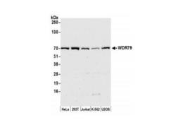
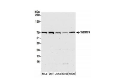
- Western Blot: WDR79 Antibody [NB100-68252] - Whole cell lysate (50 ug) from HeLa, HEK293T, Jurkat, K-562, and U2OS cells prepared using NETN lysis buffer. Antibody: Affinity purified rabbit anti-WDR79 antibody used for WB at 0.04 ug/ml. Detection: Chemiluminescence with an exposure time of 75 seconds.
- Submitted by
- Novus Biologicals (provider)
- Main image
- Experimental details
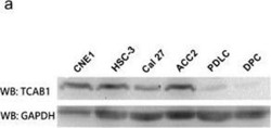
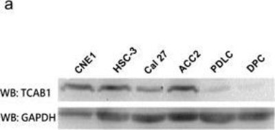
- Western Blot: WDR79 Antibody [NB100-68252] - WDR79 (TCAB1) was overexpressed in cell lines and in tissues of head and neck cancers. Protein level of WDR79 (TCAB1) in head and neck cancer cell lines (the first 4 samples) compared to human normal primary cells (the last 2 samples). Image collected and cropped by CiteAb from the following publication (http://molecular-cancer.biomedcentral.com/articles/10.1186/1476-4598-13-180), licensed under a CC-BY licence.
- Submitted by
- Novus Biologicals (provider)
- Main image

- Experimental details
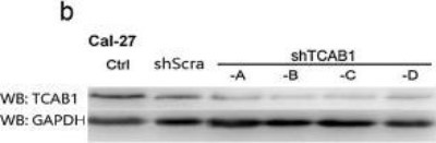
- Western Blot: WDR79 Antibody [NB100-68252] - Depletion of WDR79 (TCAB1) reduced the cell proliferation in vitro. Protein level also significantly decreased after shWDR79 (TCAB1) lentivirus treatment. Image collected and cropped by CiteAb from the following publication (http://molecular-cancer.biomedcentral.com/articles/10.1186/1476-4598-13-180), licensed under a CC-BY licence.
Supportive validation
- Submitted by
- Novus Biologicals (provider)
- Main image
- Experimental details
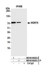
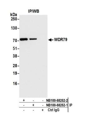
- Immunoprecipitation: WDR79 Antibody [NB100-68252] - Detection of human WDR79 by western blot of immunoprecipitates. Samples: Whole cell lysate (1.0 mg per IP reaction; 20% of IP loaded) from HeLa cells prepared using NETN lysis buffer. Antibodies: Affinity purified rabbit anti-WDR79 antibody NB100-68252 (lot NB100-68252-2) used for IP at 6 ug per reaction. WDR79 was also immunoprecipitated by a previous lot of this antibody (lot NB100-68252-1). For blotting immunoprecipitated WDR79, NB100-68252 was used at 0.04 ug/ml. Detection: Chemiluminescence with an exposure time of 10 seconds.
Supportive validation
- Submitted by
- Novus Biologicals (provider)
- Main image

- Experimental details
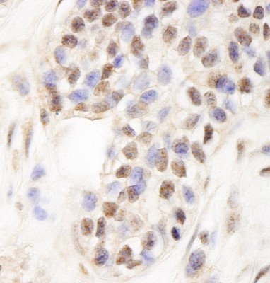
- Immunohistochemistry: WDR79 Antibody [NB100-68252] - Sample: FFPE section of human breast carcinoma. Antibody: Affinity purified rabbit anti- WDR79 used at a dilution of 1:200 (1ug/ml). Detection: DAB
- Submitted by
- Novus Biologicals (provider)
- Main image
- Experimental details
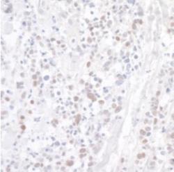
- Immunohistochemistry-Paraffin: WDR79 Antibody [NB100-68252] - Section of human lung carcinoma. Antibody: Affinity purified rabbit anti-WDR79 used at a dilution of 1:200 (1ug/ml). Detection: DAB
- Submitted by
- Novus Biologicals (provider)
- Main image
- Experimental details
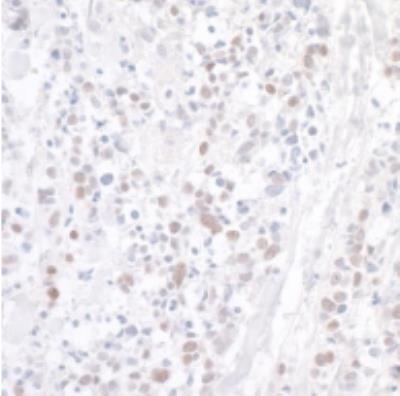
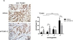
- Immunohistochemistry: WDR79 Antibody [NB100-68252] - WDR79 (TCAB1) knockdown inhibits tumor growth in vivo. Performed IHC against WDR79 (TCAB1) using mice xenografts sections. The smaller tumors expressed less WDR79 (TCAB1) compared to shScra cells. Statistical analysis of the IHC data used Aperio ImageScope software and all of the data were determined by Student's t test (*P < 0.05, **P < 0.01, ***P < 0.005). Image collected and cropped by CiteAb from the following publication (http://molecular-cancer.biomedcentral.com/articles/10.1186/1476-4598-13-180), licensed under a CC-BY licence.

- Submitted by
- Novus Biologicals (provider)
- Main image
- Experimental details
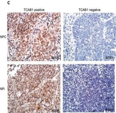
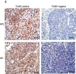
- Immunohistochemistry: WDR79 Antibody [NB100-68252] - WDR79 (TCAB1) was overexpressed in cell lines and in tissues of head and neck cancers. The typical IHC results (Left: WDR79 (TCAB1) positive, Right: WDR79 (TCAB1) negative) of WDR79 (TCAB1) in human NPC and Npi. Image collected and cropped by CiteAb from the following publication (http://molecular-cancer.biomedcentral.com/articles/10.1186/1476-4598-13-180), licensed under a CC-BY licence.