Explore
 Validate
 Learn
Learn Western blot
 Immunocytochemistry
Antibody data
- Antibody Data
- Antigen structure
- References [1]
- Comments [0]
- Validations
- Immunocytochemistry [6]
- Other assay [1]
Submit
Validation data
Reference
Comment
Report error
- Product number
- PA5-72751 - Provider product page

- Provider
- Invitrogen Antibodies
- Product name
- JMJD3 Polyclonal Antibody
- Antibody type
- Polyclonal
- Antigen
- Synthetic peptide
- Reactivity
- Human, Rat
- Host
- Rabbit
- Isotype
- IgG
- Vial size
- 100 μg
- Concentration
- 1 mg/mL
- Storage
- Store at 4°C short term. For long term storage, store at -20°C, avoiding freeze/thaw cycles.
Submitted references Activation of GPR40 attenuates neuroinflammation and improves neurological function via PAK4/CREB/KDM6B pathway in an experimental GMH rat model.
Xiao J, Cai T, Fang Y, Liu R, Flores JJ, Wang W, Gao L, Liu Y, Lu Q, Tang L, Zhang JH, Lu H, Tang J
Journal of neuroinflammation 2021 Jul 18;18(1):160
Journal of neuroinflammation 2021 Jul 18;18(1):160
No comments: Submit comment
Supportive validation
- Submitted by
- Invitrogen Antibodies (provider)
- Main image
- Experimental details
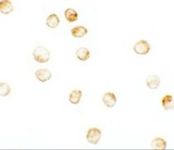
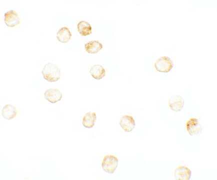
- Immunocytochemistry analysis of JMJD3 in K562 cells. The samples were probed with a JMJD3 polyclonal antibody (Product # PA5-72751) with a dilution of 2.5 µg/mL.
- Submitted by
- Invitrogen Antibodies (provider)
- Main image
- Experimental details
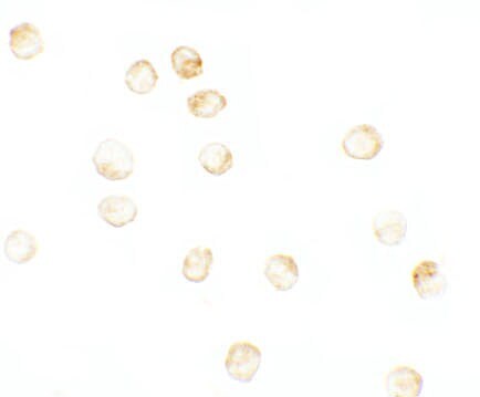
- Immunocytochemistry of K562 cells using JMJD3 Polyclonal Antibody (Product # PA5-72751) at 2.5 µg/mL. Cells were fixed with formaldehyde and blocked with 0.1 serum for 1 h at RT; antigen retrieval was by heat mediation with a citrate buffer (pH6). Samples were incubated with primary antibody overnight at 4°C. A goat anti-rabbit IgG H&L (HRP) at 1:250 was used as secondary. Counter stained with Hematoxylin.
- Submitted by
- Invitrogen Antibodies (provider)
- Main image

- Experimental details
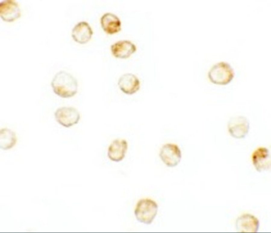
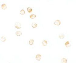
- Immunocytochemistry of K562 cells using JMJD3 Polyclonal Antibody (Product # PA5-72751) at 2.5 µg/mL. Cells were fixed with formaldehyde and blocked with 0.1 serum for 1 h at RT; antigen retrieval was by heat mediation with a citrate buffer (pH6). Samples were incubated with primary antibody overnight at 4°C. A goat anti-rabbit IgG H&L (HRP) at 1:250 was used as secondary. Counter stained with Hematoxylin.

- Submitted by
- Invitrogen Antibodies (provider)
- Main image
- Experimental details
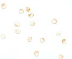
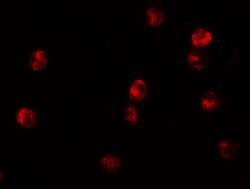
- Immunofluorescent analysis of 4% paraformaldehyde-fixed K562 cells labeling JMJD3 with JMJD3 Polyclonal Antibody (Product # PA5-72751) at 20 µg/mL, followed by goat anti-rabbit IgG secondary antibody at 1:500 dilution (red).

- Submitted by
- Invitrogen Antibodies (provider)
- Main image

- Experimental details
- Immunofluorescent analysis of 4% paraformaldehyde-fixed K562 cells labeling JMJD3 with JMJD3 Polyclonal Antibody (Product # PA5-72751) at 20 µg/mL, followed by goat anti-rabbit IgG secondary antibody at 1:500 dilution (red).

- Submitted by
- Invitrogen Antibodies (provider)
- Main image
- Experimental details
- Immunocytochemistry of K562 cells using JMJD3 Polyclonal Antibody (Product # PA5-72751) at 2.5 µg/mL. Cells were fixed with formaldehyde and blocked with 0.1 serum for 1 h at RT; antigen retrieval was by heat mediation with a citrate buffer (pH6). Samples were incubated with primary antibody overnight at 4°C. A goat anti-rabbit IgG H&L (HRP) at 1:250 was used as secondary. Counter stained with Hematoxylin.
Supportive validation
- Submitted by
- Invitrogen Antibodies (provider)
- Main image
- Experimental details
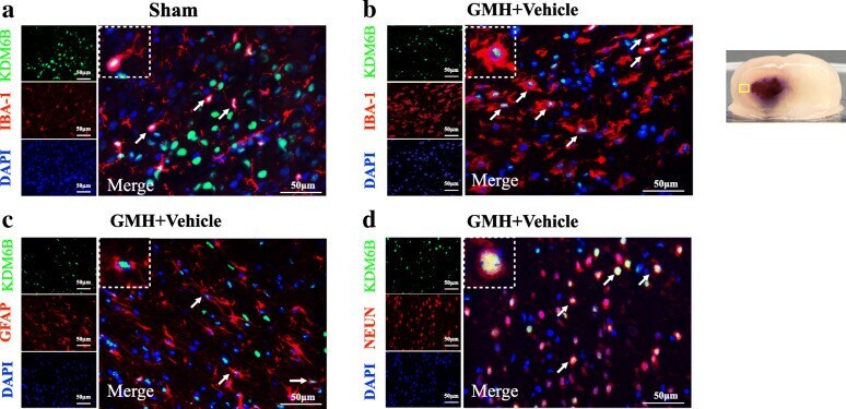
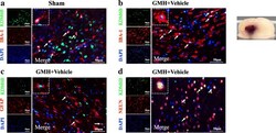
- Fig. 3 Double immunofluorescence staining of KDM6B with calcium-binding adaptor molecule 1 (Iba-1), glial fibrillary acidic protein (GFAP), and neuronal nuclei (NeuN) at 24 h after GMH. Top panel indicates the location of staining (small black box). Scale bar = 50 mum. n = 2 for each group