Explore
 Validate
 Learn
Learn Western blot
Antibody data
- Antibody Data
- Antigen structure
- References [0]
- Comments [0]
- Validations
- Western blot [2]
- Immunocytochemistry [1]
- Immunohistochemistry [6]
- Other assay [1]
Submit
Validation data
Reference
Comment
Report error
- Product number
- UM800071 - Provider product page

- Provider
- OriGene
- Proper citation
- OriGene Cat#UM800071, RRID:AB_2629181
- Product name
- ABAT mouse monoclonal antibody,clone UMAB179
- Antibody type
- Monoclonal
- Description
- ABAT mouse monoclonal antibody,clone UMAB179
- Host
- Mouse
- Conjugate
- Unconjugated
- Epitope
- ABAT
- Isotype
- IgG
- Antibody clone number
- UMAB179
- Vial size
- 100 µl
- Concentration
- 1.00mg/ml
No comments: Submit comment
Supportive validation
- Submitted by
- OriGene (provider)
- Main image
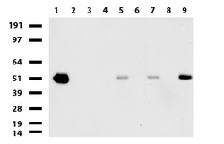
- Experimental details
- Western blot of cell lysates (35ug) from 9 different cell lines (1: HepG2, 2: HeLa, 3: SV-T2, 4: A549. 5: COS7, 6: Jurkat, 7: MDCK, 8: PC-12, 9: MCF7).
- Validation comment
- WB

- Submitted by
- OriGene (provider)
- Main image

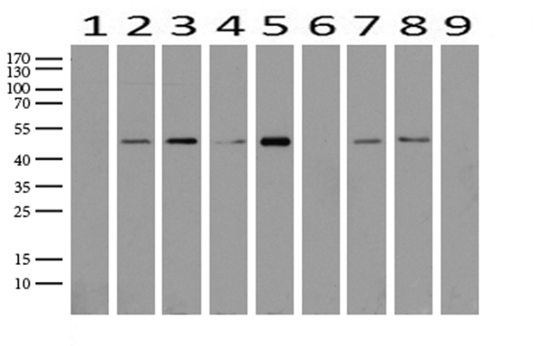
- Experimental details
- Western blot analysis of extracts (15ug) from 9 Human tissue by using anti-ABAT monoclonal antibody (1: Testis; 2: Uterus; 3: Breast; 4: Brain; 5: Liver; 6: Ovary; 7: Thyroid gland; 8: colon:;9:Spleen).(1:500) Dilution: 1:500
- Validation comment
- WB
Supportive validation
- Submitted by
- OriGene (provider)
- Main image
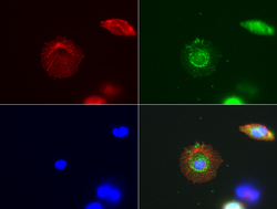
- Experimental details
- Immunofluorescent staining of HepG2 cells using anti-ABAT mouse monoclonal antibody (UM800071, green, 1:100). Actin filaments were labeled with Alexa Fluor? 594 Phalloidin (red), and nuclear with DAPI (blue).
- Validation comment
- IF

Supportive validation
- Submitted by
- OriGene (provider)
- Main image
- Experimental details
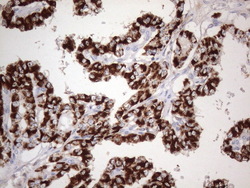
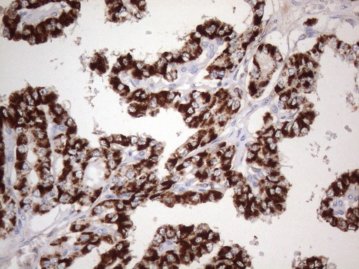
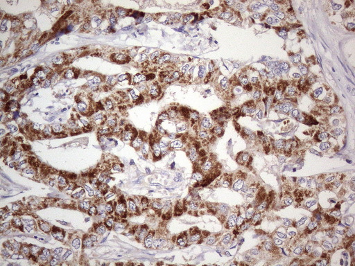
- Immunohistochemical staining of paraffin-embedded carcinoma of human thyroid tissue using ABAT clone UMAB179, mouse monoclonal antibody. Heat-induced epitope retrieval by 1mM EDTA in 10mM Tris buffer (pH8.0 ) in pressure chamber/cooker at 110C for 3 min, UM800071 was diluted 1:1000 using HRP detection and DAB chromogen. Image shows strong cytoplasmic and membranous staining is present in the tumor cells.
- Validation comment
- IHC
- Submitted by
- OriGene (provider)
- Main image
- Experimental details
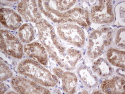
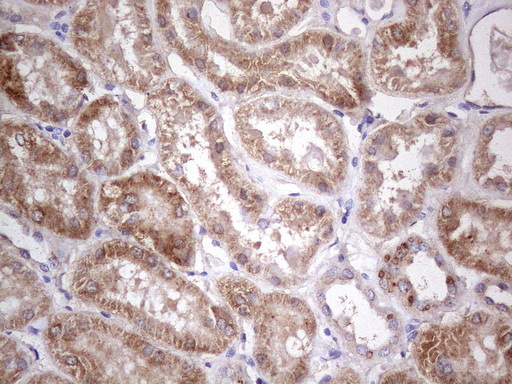
- Immunohistochemical staining of paraffin-embedded human kidney tissue using anti-ABAT mouse monoclonal antibody. Heat-induced epitope retrieval by 1mM EDTA in 10mM Tris buffer (pH8.0) in pressure chamber/cooker at 110C for 3 min, UM800071@ 1:400 shows kidney tubules with strong granular cytoplasmic staining.
- Validation comment
- IHC
- Submitted by
- OriGene (provider)
- Main image
- Experimental details
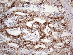
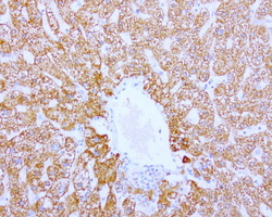
- Immunohistochemical staining of paraffin-embedded carcinoma of human liver tissue using anti-ABAT mouse monoclonal antibody.(Heat-induced epitope retrieval in pressure chamber/cooker at 110C for 3 min, UM800071(1:400). Images shows cancer cells with strong granular cytoplasmic staining.
- Validation comment
- IHC
- Submitted by
- OriGene (provider)
- Main image
- Experimental details
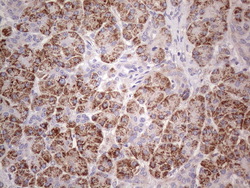
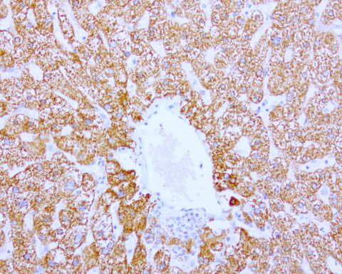
- Immunohistochemical staining of paraffin-embedded human pancreas tissue using anti-ABAT mouse monoclonal antibody. Heat-induced epitope retrieval by 1mM EDTA in 10mM Tris buffer (pH8.0) in pressure chamber/cooker at 110C for 3 min, UM800071(1:400). Images shows exocrine granular cells with strong granular cytoplasmic staining.
- Validation comment
- IHC

- Submitted by
- OriGene (provider)
- Main image
- Experimental details
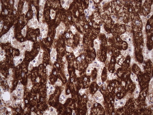
- Immunohistochemical staining of paraffin-embedded Human liver tissue using anti-ABAT mouse monoclonal antibody.(Heat-induced epitope retrieval by 1mM EDTA in 10mM Tris buffer (pH8.0) at 110C for 10 min, UM800071)(1:400)
- Validation comment
- IHC
- Submitted by
- OriGene (provider)
- Main image
- Experimental details
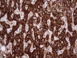
- Immunohistochemical staining of paraffin-embedded human liver using ABAT clone UMAB179, mouse monoclonal antibody at 1:400 dilution of 1mg/mL using Polink2 Broad HRP DAB for detection. UM800071 requires heat-induced epitope retrieval with citrate pH6.0 at 110C for 3 min using pressure chamber/cooker. The image shows strong cytoplasmic and membranous staining of the hepatocytes no staining in the bile duct.
- Validation comment
- IHC
Supportive validation
- Submitted by
- OriGene (provider)
- Main image
- Experimental details
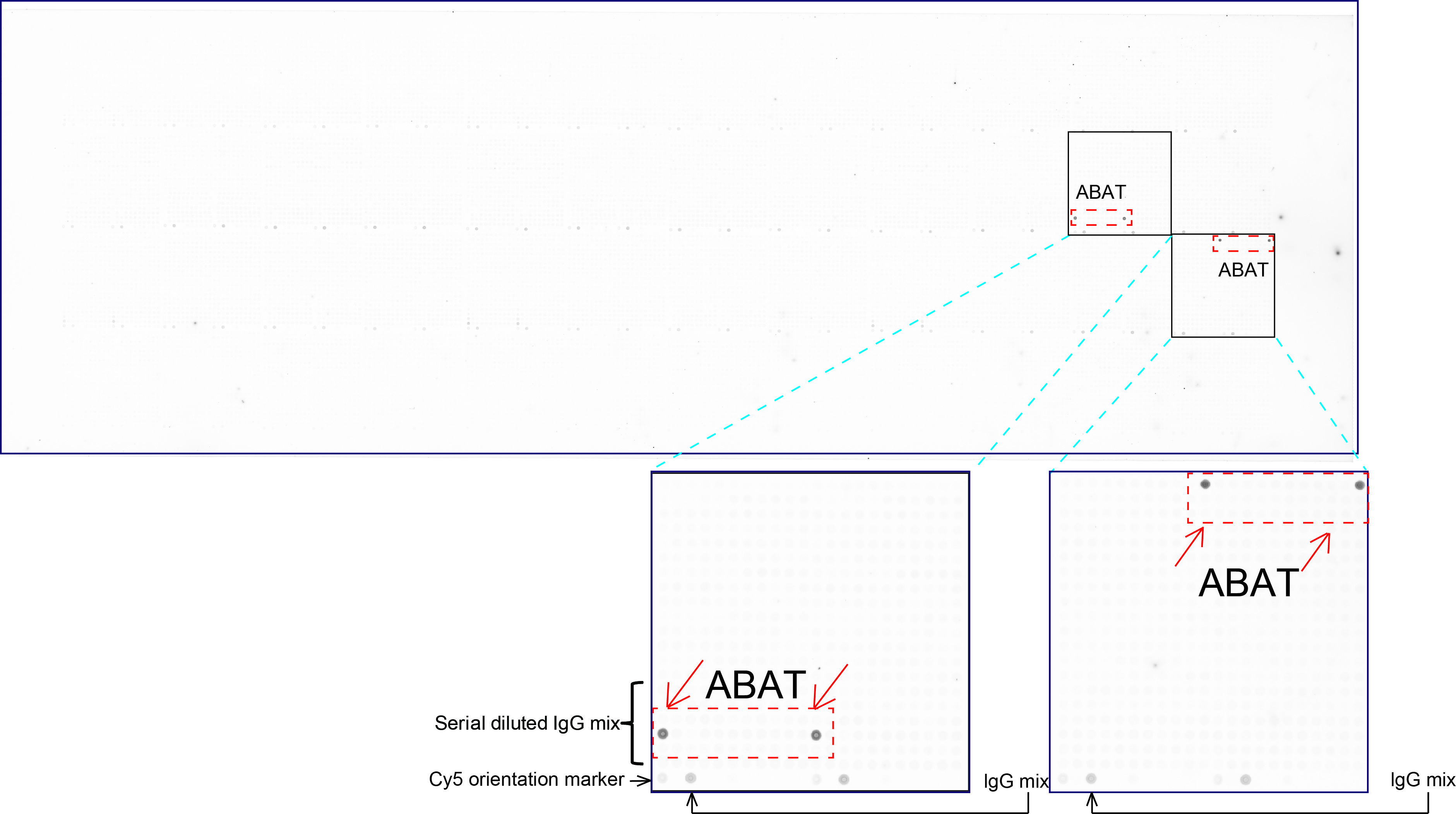
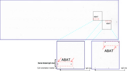
- OriGene overexpression protein microarray chip was immunostained with UltraMAB anti-ABAT mouse monoclonal antibody (UM800071). The positive reactive proteins are highlighted with two red arrows in the enlarged subarray. All the positive controls spotted in this subarray are also labeled for clarification.(1:100)
- Validation comment
- 10K-CHIP