Explore
 Validate
 Learn
Learn Western blot
 Immunocytochemistry
Antibody data
- Antibody Data
- Antigen structure
- References [1]
- Comments [0]
- Validations
- Immunocytochemistry [3]
- Immunohistochemistry [2]
Submit
Validation data
Reference
Comment
Report error
- Product number
- PA5-29829 - Provider product page

- Provider
- Invitrogen Antibodies
- Product name
- MSH3 Polyclonal Antibody
- Antibody type
- Polyclonal
- Antigen
- Recombinant full-length protein
- Description
- Recommended positive controls: A549, H1299. Predicted reactivity: Human (99%), Mouse (83%), Rat (85%). Store product as a concentrated solution. Centrifuge briefly prior to opening the vial.
- Reactivity
- Human
- Host
- Rabbit
- Isotype
- IgG
- Vial size
- 100 μL
- Concentration
- 1.79 mg/mL
- Storage
- Store at 4°C short term. For long term storage, store at -20°C, avoiding freeze/thaw cycles.
Submitted references Distinct DNA repair pathways cause genomic instability at alternative DNA structures.
McKinney JA, Wang G, Mukherjee A, Christensen L, Subramanian SHS, Zhao J, Vasquez KM
Nature communications 2020 Jan 13;11(1):236
Nature communications 2020 Jan 13;11(1):236
No comments: Submit comment
Supportive validation
- Submitted by
- Invitrogen Antibodies (provider)
- Main image
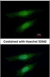
- Experimental details
- Immunofluorescent analysis of MSH3 in paraformaldehyde-fixed HeLa cells using a MSH3 polyclonal antibody (Product # PA5-29829) at a 1:500 dilution.
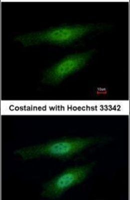
- Submitted by
- Invitrogen Antibodies (provider)
- Main image

- Experimental details
- MSH3 Polyclonal Antibody detects MSH3 protein at nucleus by immunofluorescent analysis. Sample: HeLa cells were fixed in 4% paraformaldehyde at RT for 15 min. Green: MSH3 stained by MSH3 Polyclonal Antibody (Product # PA5-29829) diluted at 1:500. Blue: Fluoroshield with DAPI . Scale bar= 10 µm.
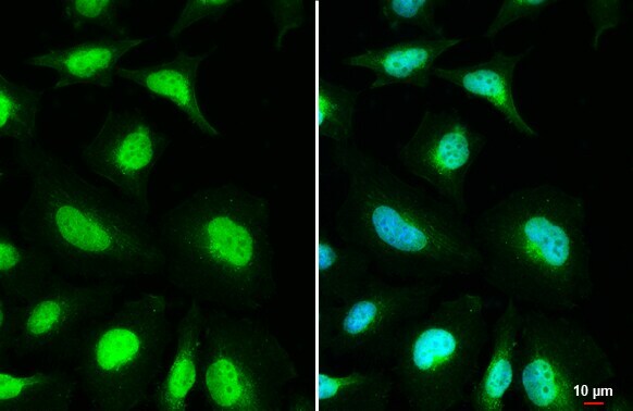

Supportive validation
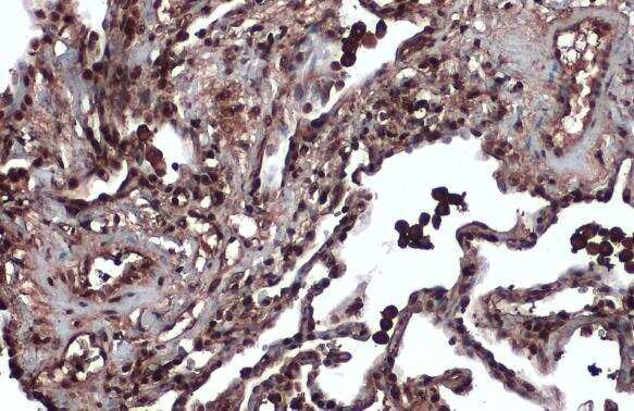
- Submitted by
- Invitrogen Antibodies (provider)
- Main image
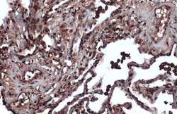
- Experimental details
- Immunohistochemistry (Paraffin) analysis of MSH3 was performed in paraffin-embedded human lung cancer tissue using MSH3 Polyclonal Antibody (Product # PA5-29829) at a dilution of 1:600. Antigen Retrieval: Citrate buffer, pH 6.0, 15 min.
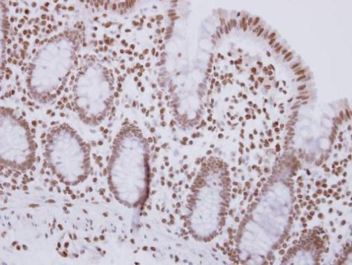
- Submitted by
- Invitrogen Antibodies (provider)
- Main image
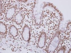
- Experimental details
- Immunohistochemical analysis of paraffin-embedded human colon carcinoma, using MSH3 (Product # PA5-29829) antibody at 1:500 dilution. Antigen Retrieval: EDTA based buffer, pH 8.0, 15 min.