 Explore
 Validate
 Learn
Learn Western blot
Antibody data
- Antibody Data
- Antigen structure
- References [0]
- Comments [0]
- Validations
- Western blot [7]
- Immunohistochemistry [3]
Submit
Validation data
Reference
Comment
Report error
- Product number
- PA5-29719 - Provider product page

- Provider
- Invitrogen Antibodies
- Product name
- ATP1B1 Polyclonal Antibody
- Antibody type
- Polyclonal
- Antigen
- Recombinant protein fragment
- Description
- Recommended positive controls: K562, HL-60, mouse brain, Rat brain. Predicted reactivity: Mouse (90%), Rat (92%), Dog (91%), Pig (89%), Rabbit (87%), Sheep (91%), Rhesus Monkey (96%), Chimpanzee (99%), Guinea pig (86%). Store product as a concentrated solution. Centrifuge briefly prior to opening the vial.
- Reactivity
- Human, Mouse, Rat
- Host
- Rabbit
- Isotype
- IgG
- Vial size
- 100 µL
- Concentration
- 1 mg/mL
- Storage
- Store at 4°C short term. For long term storage, store at -20°C, avoiding freeze/thaw cycles.
No comments: Submit comment
Supportive validation
- Submitted by
- Invitrogen Antibodies (provider)
- Main image
- Experimental details
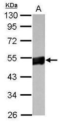
- Western blot analysis of Sodium Potassium ATPase Beta 1 using 50 µg of mouse brain lysate. Samples were loaded onto a 10% SDS-PAGE gel and probed with a Sodium Potassium ATPase Beta 1 polyclonal antibody (Product # PA5-29719) at a dilution of 1:10,000.

- Submitted by
- Invitrogen Antibodies (provider)
- Main image
- Experimental details
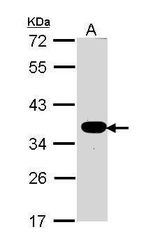
- Western blot analysis of Sodium Potassium ATPase Beta 1 using 30 µg of MOLT4 lysate. Samples were loaded onto a 10% SDS-PAGE gel and probed with a Sodium Potassium ATPase Beta 1 polyclonal antibody (Product # PA5-29719) at a dilution of 1:1000.
- Submitted by
- Invitrogen Antibodies (provider)
- Main image
- Experimental details
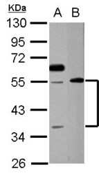
- Western Blot analysis of ATP1B1 was performed by separating 30 µg of various whole cell extracts by 10% SDS PAGE. Proteins were transferred to a membrane and probed with a ATP1B1 Polyclonal Antibody (Product # PA5-29719) at a dilution of 1:1000. A: K562, B: HL-60.

- Submitted by
- Invitrogen Antibodies (provider)
- Main image
- Experimental details
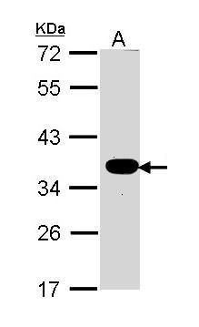
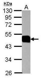
- Western Blot using ATP1B1 Polyclonal Antibody (Product # PA5-29719). Sample (50 µg of whole cell lysate). Lane A: Rat brain. 10% SDS PAGE. ATP1B1 Polyclonal Antibody (Product # PA5-29719) diluted at 1:1,000. The HRP-conjugated anti-rabbit IgG antibody was used to detect the primary antibody.

- Submitted by
- Invitrogen Antibodies (provider)
- Main image
- Experimental details
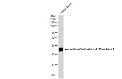
- Western Blot using ATP1B1 Polyclonal Antibody (Product # PA5-29719). Human brain (30 µg) was separated by 10% SDS-PAGE, and the membrane was blotted with ATP1B1 Polyclonal Antibody (Product # PA5-29719) diluted at 1:1,000. The HRP-conjugated anti-rabbit IgG antibody was used to detect the primary antibody.

- Submitted by
- Invitrogen Antibodies (provider)
- Main image
- Experimental details
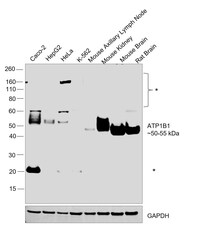
- Western blot was performed using Anti-ATP1B1 Polyclonal Antibody (Product # PA5-29719) and a 50-55 kDa band corresponding to ATP1B1 was observed across cell lines and tissue tested but not in K-562 and Mouse Lymph Node which are reported to be low or negative for ATP1B1 expression. An additional uncharacterized band (*) was observed in a few models. Whole cell extracts (30 µg lysate) of Caco-2 (Lane 1), Hep G2 (Lane 2), HeLa (Lane 3), K-562 (Lane 4), Mouse Axillary Lymph node (Lane 5), Mouse Kidney (Lane 6), Mouse Brain (Lane 7), Rat Brain (Lane 8) were electrophoresed using NuPAGE™ 4-12% Bis-Tris Protein Gel (Product # NP0322BOX). Resolved proteins were then transferred onto a nitrocellulose membrane (Product # IB23002) by iBlot® 2 Dry Blotting System (Product # IB21001). The blot was probed with the primary antibody (1:1000 dilution) and detected by chemiluminescence with Goat anti-Rabbit IgG (H+L) Superclonal™ Recombinant Secondary Antibody, HRP (Product # A27036,1:20,000 dilution) using the iBright™ FL1500 Imaging System (Product # A44115). Chemiluminescent detection was performed using SuperSignal™ West Pico PLUS Chemiluminescent Substrate (Product # 34580).

- Submitted by
- Invitrogen Antibodies (provider)
- Main image

- Experimental details
- Western Blot analysis of ATP1B1 was performed by separating 50 µg of mouse brain lysates by 10% SDS PAGE. Proteins were transferred to a membrane and probed with a ATP1B1 Polyclonal Antibody (Product # PA5-29719) at a dilution of 1:1000.

Supportive validation
- Submitted by
- Invitrogen Antibodies (provider)
- Main image
- Experimental details
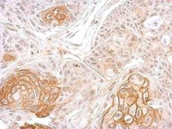
- Immunohistochemical analysis of paraffin-embedded MDAMB468 xenograft, using ATPase beta1 (Na+/K+) (Product # PA5-29719) antibody at 1:100 dilution. Antigen Retrieval: EDTA based buffer, pH 8.0, 15 min.
- Submitted by
- Invitrogen Antibodies (provider)
- Main image
- Experimental details
- Sodium Potassium ATPase Beta 1 antibody detects ATP1B1 protein at membrane on Cal27 xenograft by immunohistochemical analysis. Sample: Paraffin-embedded Cal27 xenograft. Sodium Potassium ATPase Beta 1 antibody (Product # PA5-29719) dilution: 1:500. Antigen Retrieval: EDTA based buffer, pH 8.0, 15 min.

- Submitted by
- Invitrogen Antibodies (provider)
- Main image
- Experimental details
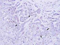
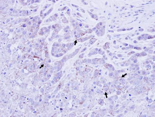
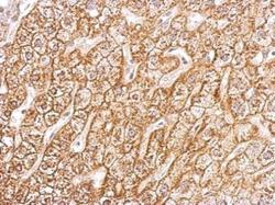
- Sodium Potassium ATPase Beta 1 antibody detects ATP1B1 protein at membrane on human hepatoma by immunohistochemical analysis. Sample: Paraffin-embedded hepatoma tissue. Sodium Potassium ATPase Beta 1 antibody (Product # PA5-29719) dilution: 1:500. Antigen Retrieval: EDTA based buffer, pH 8.0, 15 min.
