 Explore
 Validate
 Learn
Learn Western blot
 ELISA
ELISAAntibody data
- Antibody Data
- Antigen structure
- References [0]
- Comments [0]
- Validations
- Western blot [2]
- Immunocytochemistry [2]
- Immunohistochemistry [3]
- Flow cytometry [3]
Submit
Validation data
Reference
Comment
Report error
- Product number
- MA5-29000 - Provider product page

- Provider
- Invitrogen Antibodies
- Product name
- ADAM15 Recombinant Rabbit Monoclonal Antibody (007)
- Antibody type
- Monoclonal
- Antigen
- Recombinant full-length protein
- Description
- This product is preservative free. It is recommended to add sodium azide to avoid contamination (final concentration 0.05%-0.1%). Recombinant rabbit monoclonal antibodies are produced using in vitro expression systems. The expression systems are developed by cloning in the specific antibody DNA sequences from immunoreactive rabbits. Then, individual clones are screened to select the best candidates for production. The advantages of using recombinant rabbit monoclonal antibodies include: better specificity and sensitivity, lot-to-lot consistency, animal origin-free formulations, and broader immunoreactivity to diverse targets due to larger rabbit immune repertoire. This antibody has specificity for Human ADAM15/MDC15.
- Reactivity
- Human
- Host
- Rabbit
- Isotype
- IgG
- Antibody clone number
- 7
- Vial size
- 100 μL
- Concentration
- 1.33 mg/mL
- Storage
- Store at 4°C short term. For long term storage, store at -20°C, avoiding freeze/thaw cycles.
No comments: Submit comment
Supportive validation
- Submitted by
- Invitrogen Antibodies (provider)
- Main image
- Experimental details
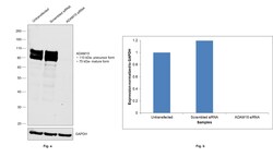
- Knockdown of ADAM15 was achieved by transfecting PC-3 with ADAM15 specific siRNAs (Silencer® select Product # s16683, s16682). Western blot analysis (Fig. a) was performed using Whole cell extracts from the ADAM15 knockdown cells (lane 3), non-targeting scrambled siRNA transfected cells (lane 2) and untransfected cells (lane 1). The blot was probed with ADAM15 Recombinant Rabbit Monoclonal Antibody (7) (Product # MA5-29000, 1:1,000) and Goat anti-Rabbit IgG (H+L) Superclonal™ Recombinant Secondary Antibody, HRP (Product # A27036, 1:20,000). Densitometric analysis of this western blot is shown in histogram (Fig. b). Decrease in signal upon siRNA mediated knock down confirms that antibody is specific to ADAM15.

- Submitted by
- Invitrogen Antibodies (provider)
- Main image
- Experimental details
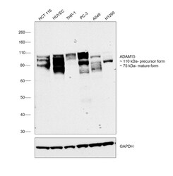
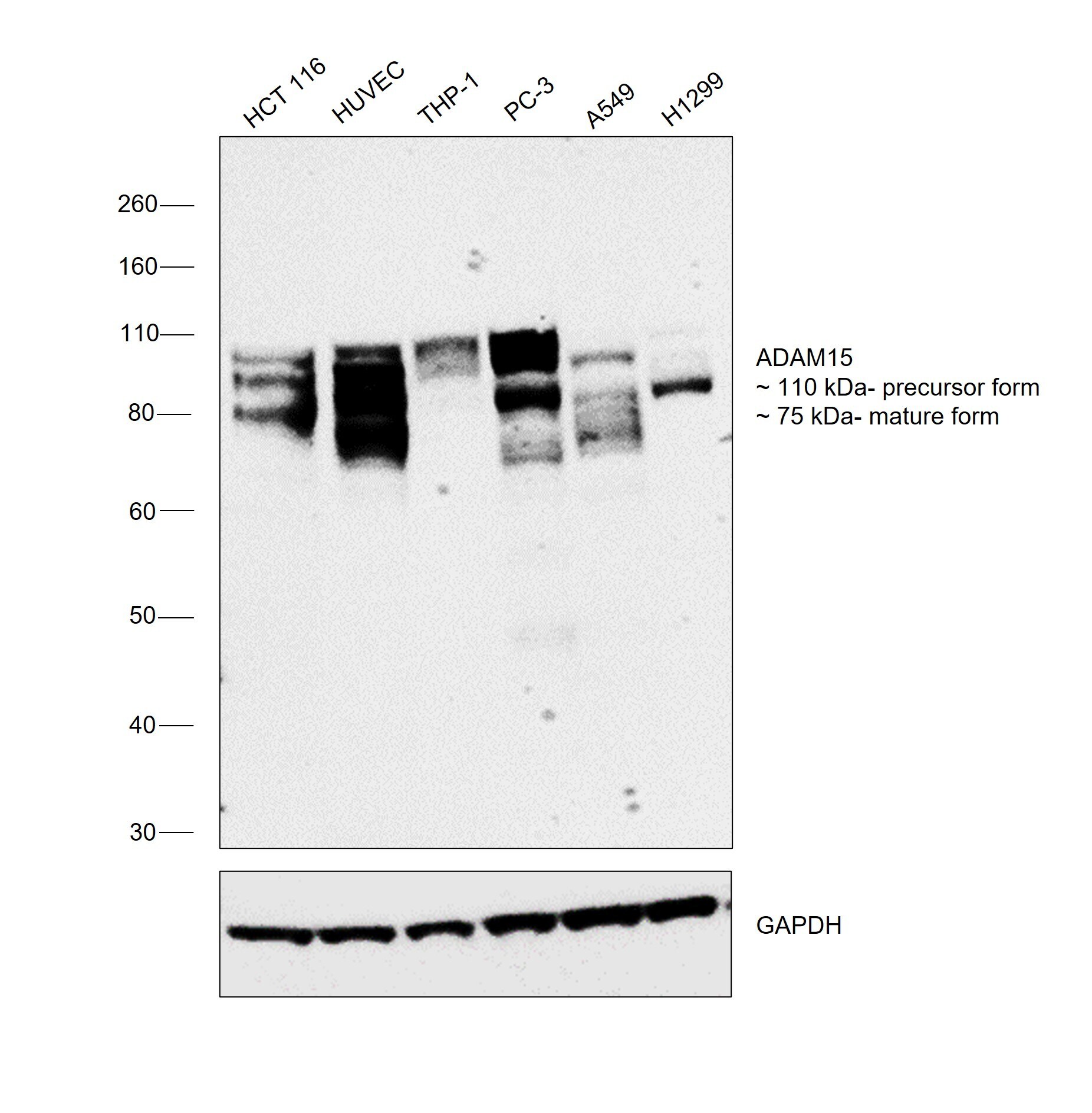
- Western blot was performed using ADAM15 Recombinant Rabbit Monoclonal Antibody (7) (Product # MA5-29000) and a 110 kDa band corresponding to precursor form and a 75 kDa band corresponding to mature form of ADAM15 was observed across cell lines. Whole cell extracts (30 µg lysate) of HCT 116 (Lane 1), HUVEC (Lane 2), THP-1 (Lane 3), PC-3 (Lane 4), A549 (Lane 5), H1299 (Lane 6) were electrophoresed using NuPAGE™ 4-12% Bis-Tris Protein Gel (Product # NP0321BOX), 10 well. Resolved proteins were then transferred onto a nitrocellulose membrane (Product # IB33001) by iBlot™ 3 Western Blot Transfer Device (Product # IB31001). The blot was probed with the primary antibody (1:1,000) and detected by chemiluminescence with Goat anti-Rabbit IgG (H+L) Superclonal™ Recombinant Secondary Antibody, HRP (Product # A27036, 1:10,000) using the iBright™ FL1500 Imaging System (Product # A44115). Chemiluminescent detection was performed using SuperSignal™ West Pico PLUS Chemiluminescent Substrate (Product # 34580).
Supportive validation
- Submitted by
- Invitrogen Antibodies (provider)
- Main image

- Experimental details
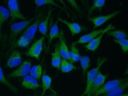
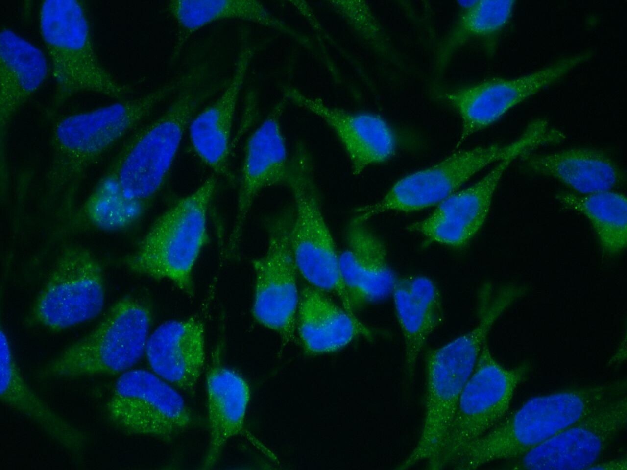
- Immunofluorescence staining of Human ADAM15 in Hela cells. Cells were fixed with 4% PFA, permeabilzed with 1% Triton X-100 in PBS, blocked with 10% serum, and incubated with ADAM15 Recombinant Rabbit Monoclonal Antibody (7) (Product # MA5-29000, 1:60) at 37°C 1 hour. Then cells were stained with the Alexa Fluor® 488-conjugated Goat Anti-rabbit IgG secondary antibody (green). Positive staining was localized to cytoplasm.

- Submitted by
- Invitrogen Antibodies (provider)
- Main image
- Experimental details
- Immunofluorescence staining of Human ADAM15 in Hela cells. Cells were fixed with 4% PFA, permeabilzed with 1% Triton X-100 in PBS, blocked with 10% serum, and incubated with ADAM15 Recombinant Rabbit Monoclonal Antibody (7) (Product # MA5-29000, 1:60) at 37°C 1 hour. Then cells were stained with the Alexa Fluor® 488-conjugated Goat Anti-rabbit IgG secondary antibody (green). Positive staining was localized to cytoplasm.
Supportive validation
- Submitted by
- Invitrogen Antibodies (provider)
- Main image

- Experimental details
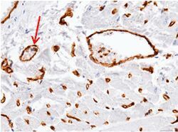
- Immunohistochemical staining of formalin fixed, paraffin-embedded human heart showing blood vessel staining (red arrow) using ADAM15 Recombinant Rabbit Monoclonal Antibody (7) (Product # MA5-29000, 1:200).

- Submitted by
- Invitrogen Antibodies (provider)
- Main image
- Experimental details
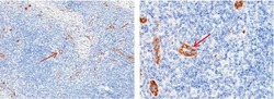
- Immunohistochemical staining of formalin fixed, paraffin-embedded human lymph node showing membrane staining of endothelial cells (red arrow) using ADAM15 Recombinant Rabbit Monoclonal Antibody (7) (Product # MA5-29000, 1:2,000).

- Submitted by
- Invitrogen Antibodies (provider)
- Main image
- Experimental details
- Immunohistochemical staining of formalin fixed, paraffin-embedded human heart showing blood vessel staining (red arrow) using ADAM15 Recombinant Rabbit Monoclonal Antibody (7) (Product # MA5-29000, 1:200).

Supportive validation
- Submitted by
- Invitrogen Antibodies (provider)
- Main image
- Experimental details
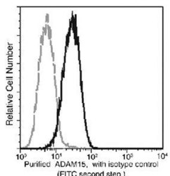
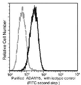
- Profile of anti-ADAM15 reactivity on MCF-7 cells analyzed by flow cytometry. Cells were stained with an ADAM15 Monoclonal Antibody (Product # MA5-29000)

- Submitted by
- Invitrogen Antibodies (provider)
- Main image

- Experimental details
- Profile of ADAM15 on MCF-7 cells analyzed by flow cytometry using ADAM15 Recombinant Rabbit Monoclonal Antibody (7) (Product # MA5-29000).
- Submitted by
- Invitrogen Antibodies (provider)
- Main image

- Experimental details
- Profile of ADAM15 on MCF-7 cells analyzed by flow cytometry using ADAM15 Recombinant Rabbit Monoclonal Antibody (7) (Product # MA5-29000).
