Explore
 Validate
 Learn
Learn Western blot
 Immunocytochemistry
Antibody data
- Antibody Data
- Antigen structure
- References [7]
- Comments [0]
- Validations
- Immunocytochemistry [1]
- Immunohistochemistry [1]
Submit
Validation data
Reference
Comment
Report error
- Product number
- HPA000962 - Provider product page

- Provider
- Atlas Antibodies
- Proper citation
- Atlas Antibodies Cat#HPA000962, RRID:AB_1079722
- Product name
- Anti-PYGL
- Antibody type
- Polyclonal
- Description
- Polyclonal Antibody against Human PYGL, Gene description: phosphorylase, glycogen, liver, Validated applications: ICC, WB, IHC, Uniprot ID: P06737, Storage: Store at +4°C for short term storage. Long time storage is recommended at -20°C.
- Reactivity
- Human
- Host
- Rabbit
- Conjugate
- Unconjugated
- Isotype
- IgG
- Vial size
- 100 µl
- Concentration
- 0.1 mg/ml
- Storage
- Store at +4°C for short term storage. Long time storage is recommended at -20°C.
- Handling
- The antibody solution should be gently mixed before use.
Submitted references Integrated genomic and proteomic analyses identify PYGL as a novel experimental therapeutic target for clear cell renal cell carcinoma
Deep topographic proteomics of a human brain tumour.
Identification of Potential Muscle Biomarkers in McArdle Disease: Insights from Muscle Proteome Analysis
Liver glycogen phosphorylase is upregulated in glioblastoma and provides a metabolic vulnerability to high dose radiation
Glycogen metabolism is dispensable for tumour progression in clear cell renal cell carcinoma
AMPK antagonizes hepatic glucagon-stimulated cyclic AMP signalling via phosphorylation-induced activation of cyclic nucleotide phosphodiesterase 4B
Expression of Glycogen Phosphorylase Isoforms in Cultured Muscle from Patients with McArdle's Disease Carrying the p.R771PfsX33 PYGM Mutation
Li M, Zhu G, Liu Y, Li X, Zhou Y, Li C, Wang M, Zhang J, Wang Z, Tan S, Chen W, Zhang H
Heliyon 2024;10(6):e28295
Heliyon 2024;10(6):e28295
Deep topographic proteomics of a human brain tumour.
Davis S, Scott C, Oetjen J, Charles PD, Kessler BM, Ansorge O, Fischer R
Nature communications 2023 Nov 24;14(1):7710
Nature communications 2023 Nov 24;14(1):7710
Identification of Potential Muscle Biomarkers in McArdle Disease: Insights from Muscle Proteome Analysis
García-Consuegra I, Asensio-Peña S, Garrido-Moraga R, Pinós T, Domínguez-González C, Santalla A, Nogales-Gadea G, Serrano-Lorenzo P, Andreu A, Arenas J, Zugaza J, Lucia A, Martín M
International Journal of Molecular Sciences 2022;23(9):4650
International Journal of Molecular Sciences 2022;23(9):4650
Liver glycogen phosphorylase is upregulated in glioblastoma and provides a metabolic vulnerability to high dose radiation
Zois C, Hendriks A, Haider S, Pires E, Bridges E, Kalamida D, Voukantsis D, Lagerholm B, Fehrmann R, den Dunnen W, Tarasov A, Baba O, Morris J, Buffa F, McCullagh J, Jalving M, Harris A
Cell Death & Disease 2022;13(6)
Cell Death & Disease 2022;13(6)
Glycogen metabolism is dispensable for tumour progression in clear cell renal cell carcinoma
Xie H, Song J, Godfrey J, Riscal R, Skuli N, Nissim I, Simon M
Nature Metabolism 2021;3(3):327-336
Nature Metabolism 2021;3(3):327-336
AMPK antagonizes hepatic glucagon-stimulated cyclic AMP signalling via phosphorylation-induced activation of cyclic nucleotide phosphodiesterase 4B
Johanns M, Lai Y, Hsu M, Jacobs R, Vertommen D, Van Sande J, Dumont J, Woods A, Carling D, Hue L, Viollet B, Foretz M, Rider M
Nature Communications 2016;7(1)
Nature Communications 2016;7(1)
Expression of Glycogen Phosphorylase Isoforms in Cultured Muscle from Patients with McArdle's Disease Carrying the p.R771PfsX33 PYGM Mutation
Palau F, Nogales-Gadea G, Mormeneo E, García-Consuegra I, Rubio J, Orozco A, Arenas J, Martín M, Lucia A, Gómez-Foix A, Martí R, Andreu A
PLoS ONE 2010;5(10):e13164
PLoS ONE 2010;5(10):e13164
No comments: Submit comment
Supportive validation
- Submitted by
- Atlas Antibodies (provider)
- Main image
- Experimental details
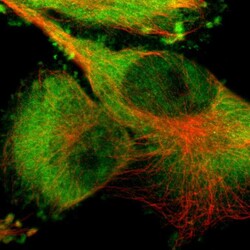
- Immunofluorescent staining of human cell line U-251 MG shows localization to nucleoplasm, plasma membrane & cytosol.
- Sample type
- Human

Supportive validation
- Submitted by
- Atlas Antibodies (provider)
- Enhanced method
- Orthogonal validation
- Main image
- Experimental details
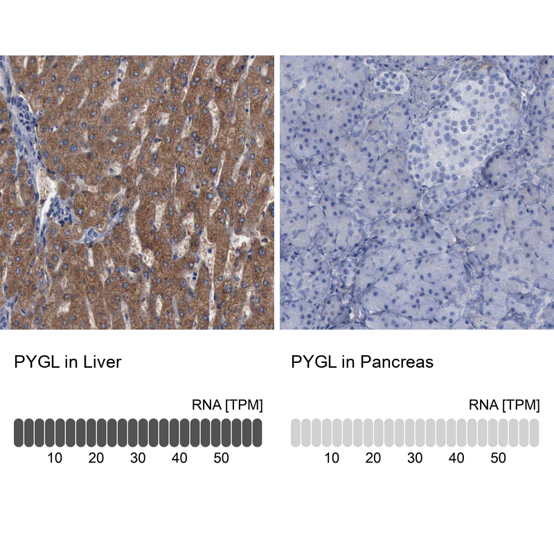
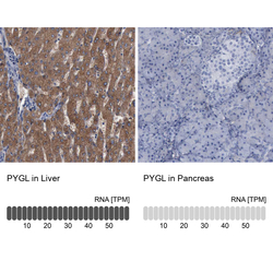
- Immunohistochemistry analysis in human liver and pancreas tissues using HPA000962 antibody. Corresponding PYGL RNA-seq data are presented for the same tissues.
- Sample type
- Human
- Protocol
- Protocol