Explore
 Validate
 Learn
Learn Western blot
 Immunocytochemistry
Antibody data
- Antibody Data
- Antigen structure
- References [3]
- Comments [0]
- Validations
- Immunocytochemistry [1]
- Immunohistochemistry [1]
Submit
Validation data
Reference
Comment
Report error
- Product number
- HPA004119 - Provider product page

- Provider
- Atlas Antibodies
- Proper citation
- Atlas Antibodies Cat#HPA004119, RRID:AB_1079723
- Product name
- Anti-PYGL
- Antibody type
- Polyclonal
- Description
- Polyclonal Antibody against Human PYGL, Gene description: phosphorylase, glycogen, liver, Validated applications: WB, IHC, ICC, Uniprot ID: P06737, Storage: Store at +4°C for short term storage. Long time storage is recommended at -20°C.
- Reactivity
- Human
- Host
- Rabbit
- Conjugate
- Unconjugated
- Isotype
- IgG
- Vial size
- 100 µl
- Concentration
- 0.2 mg/ml
- Storage
- Store at +4°C for short term storage. Long time storage is recommended at -20°C.
- Handling
- The antibody solution should be gently mixed before use.
Submitted references Integrated genomic and proteomic analyses identify PYGL as a novel experimental therapeutic target for clear cell renal cell carcinoma
BAP1 Loss Is Associated with Higher ASS1 Expression in Epithelioid Mesothelioma: Implications for Therapeutic Stratification
Metabolic enzymes in glial cells of the honeybee brain and their associations with aging, starvation and food response
Li M, Zhu G, Liu Y, Li X, Zhou Y, Li C, Wang M, Zhang J, Wang Z, Tan S, Chen W, Zhang H
Heliyon 2024;10(6):e28295
Heliyon 2024;10(6):e28295
BAP1 Loss Is Associated with Higher ASS1 Expression in Epithelioid Mesothelioma: Implications for Therapeutic Stratification
Barnett S, Kenyani J, Tripari M, Butt Z, Grosman R, Querques F, Shaw L, Silva L, Goate Z, Marciniak S, Rassl D, Jackson R, Lian L, Szlosarek P, Sacco J, Coulson J
Molecular Cancer Research 2023;21(5):411-427
Molecular Cancer Research 2023;21(5):411-427
Metabolic enzymes in glial cells of the honeybee brain and their associations with aging, starvation and food response
Gronenberg W, Shah A, Kreibich C, Amdam G, Münch D
PLOS ONE 2018;13(6):e0198322
PLOS ONE 2018;13(6):e0198322
No comments: Submit comment
Supportive validation
- Submitted by
- Atlas Antibodies (provider)
- Main image
- Experimental details
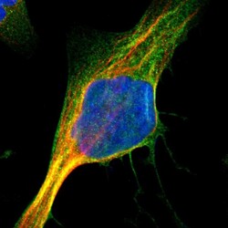
- Immunofluorescent staining of human cell line U-2 OS shows localization to plasma membrane & cytosol.
- Sample type
- Human

Supportive validation
- Submitted by
- Atlas Antibodies (provider)
- Enhanced method
- Orthogonal validation
- Main image
- Experimental details
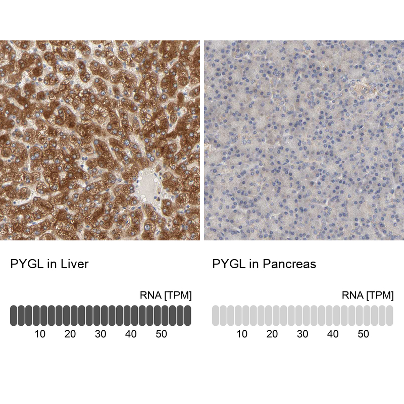
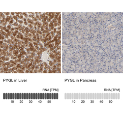
- Immunohistochemistry analysis in human liver and pancreas tissues using HPA004119 antibody. Corresponding PYGL RNA-seq data are presented for the same tissues.
- Sample type
- Human
- Protocol
- Protocol