Explore
 Validate
 Learn
Learn Western blot
 Other assay
Antibody data
- Antibody Data
- Antigen structure
- References [1]
- Comments [0]
- Validations
- Other assay [1]
Submit
Validation data
Reference
Comment
Report error
- Product number
- PA5-48853 - Provider product page

- Provider
- Invitrogen Antibodies
- Product name
- FUNDC1 Polyclonal Antibody
- Antibody type
- Polyclonal
- Antigen
- Synthetic peptide
- Description
- Predicted to react with bovine, mouse and rat based on sequence homology.
- Reactivity
- Human, Mouse
- Host
- Rabbit
- Isotype
- IgG
- Vial size
- 400 μL
- Concentration
- 0.45 mg/mL
- Storage
- Store at 4°C short term. For long term storage, store at -20°C, avoiding freeze/thaw cycles.
Submitted references MARCH5 mediates NOXA-dependent MCL1 degradation driven by kinase inhibitors and integrated stress response activation.
Arai S, Varkaris A, Nouri M, Chen S, Xie L, Balk SP
eLife 2020 Jun 2;9
eLife 2020 Jun 2;9
No comments: Submit comment
Supportive validation
- Submitted by
- Invitrogen Antibodies (provider)
- Main image
- Experimental details
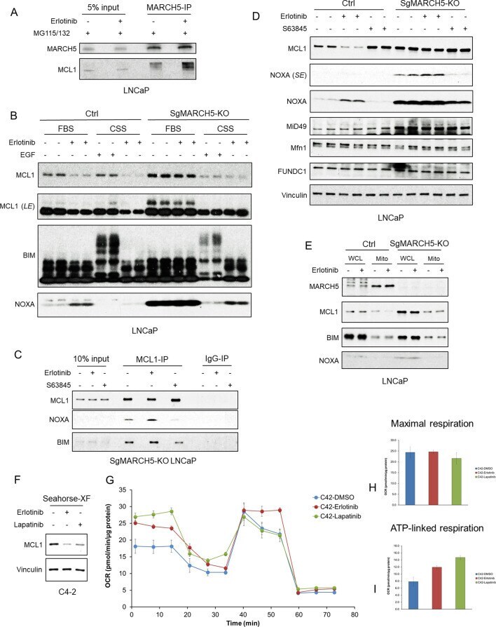
- Figure 4. EGFR inhibition enhances MARCH5-MCL1 interaction without altering MARCH5 activity. ( A ) LNCaP cells were pretreated with proteasome inhibitors MG115 (10 muM) and MG132 (10 muM) for 1 hr, followed by treatment with erlotinib (10 muM) or DMSO for 3 hr. The cell lysates were subject to immunoprecipitation using anti-MARCH5 rabbit antibody with protein A agarose, followed by immunoblotting with anti-MARCH5 rabbit antibody or anti-MCL1 mouse antibody. The decreased mobility of MCL1 in the IP lanes may reflect impaired migration due to large amounts of Ig in the sample, but phosphorylation or other posttranslational modification is possible. ( B ) SgMARCH5-KO or control LNCaP cells were pre-incubated in normal serum medium (FBS) or charcoal-stripped serum medium (CSS) for 1 day, followed by treatment with erlotinib (10 muM) for 3 hr or EGF (100 ng/ml) for 30 min. LE, long exposure. ( C ) SgMARCH5-KO LNCaP cells were treated with erlotinib (10 muM), S63845 (0.5 muM), or DMSO for 3 hr. The cell lysates were immunopurified with anti-MCL1 rabbit antibody or control rabbit IgG and protein A agarose, followed by immunoblotting with mouse antibodies targeting for indicated proteins. ( D ) SgMARCH5-KO or control LNCaP cells were treated with erlotinib (10 muM), S63845 (500 nM), or DMSO for 3 hr. SE, short exposure. ( E ) SgMARCH5-KO or control LNCaP cells were treated with erlotinib (10 muM) for 2 hr. Proteins extracted from whole cell lysates (WCL) or isolated mitochondria (Mit