Explore
 Validate
 Learn
Learn Western blot
Antibody data
- Antibody Data
- Antigen structure
- References [1]
- Comments [0]
- Validations
- Western blot [2]
- Immunocytochemistry [2]
- Other assay [1]
Submit
Validation data
Reference
Comment
Report error
- Product number
- PA1-1070 - Provider product page

- Provider
- Invitrogen Antibodies
- Product name
- Ghrelin Polyclonal Antibody
- Antibody type
- Polyclonal
- Antigen
- Synthetic peptide
- Description
- PA1-1070 detects Obestatin in mouse and rat samples. This product does not detect endogenous levels of the hormone in rat stomach. PA1-1070 has been successfully used in Western blot procedures. By Western blot, this antibody detects a ~2.5 kDa protein representing synthetic Obestatin. The PA1-1070 immunogen is a synthetic peptide corresponding to residues F(1) N A P F D V G I K L S G A Q Y Q Q H G R A L(23) of the rat Obestatin gene. This sequence is conserved in mouse and only one amino acid residue difference in human. This peptide (Cat. # PEP-290) is available for use in neutralization and control experiments.
- Reactivity
- Human, Mouse, Rat
- Host
- Rabbit
- Isotype
- IgG
- Vial size
- 100 μg
- Concentration
- 1 mg/mL
- Storage
- -20°C, Avoid Freeze/Thaw Cycles
Submitted references Study of the Ghrelin/LEAP-2 Ratio in Humans and Rats during Different Phases of Pregnancy.
Garcés MF, Buell-Acosta JD, Ángel-Müller E, Parada-Baños AJ, Acosta-Alvarez J, Saavedra-López HF, Franco-Vega R, Maldonado-Acosta LM, Escobar-Cordoba F, Vásquez-Romero K, Lacunza E, Caminos-Cepeda SA, Nogueiras R, Diéguez C, Ruiz-Parra AI, Caminos JE
International journal of molecular sciences 2022 Aug 23;23(17)
International journal of molecular sciences 2022 Aug 23;23(17)
No comments: Submit comment
Supportive validation
- Submitted by
- Invitrogen Antibodies (provider)
- Main image
- Experimental details
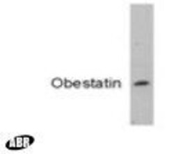
- Western blot detection of Obestatin using Product # PA1-1070.

- Submitted by
- Invitrogen Antibodies (provider)
- Main image
- Experimental details
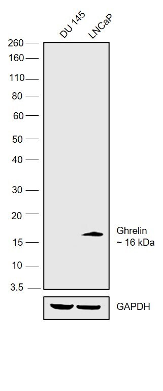
- Western blot was performed using Anti-Ghrelin Polyclonal Antibody (Product # PA1-1070) and a 16 kDa band corresponding to Ghrelin was observed in LNCaP but not in DU 145. Whole cell extracts (30 µg lysate) of DU 145 (Lane 1), LNCaP (Lane 2) were electrophoresed using NuPAGE™ 12% Bis-Tris Protein Gel (Product # NP0341BOX). Resolved proteins were then transferred onto a Nitrocellulose membrane (Product # IB23001) by iBlot® 2 Dry Blotting System (Product # IB21001). The blot was probed with the primary antibody (1:1000) and detected by chemiluminescence with Goat anti-Rabbit IgG (Heavy Chain) Superclonal™ Recombinant Secondary Antibody, HRP (Product # A27036, 1:4000) using the iBright FL 1000 (Product # A32752). Chemiluminescent detection was performed using Novex® ECL Chemiluminescent Substrate Reagent Kit (Product # WP20005).
Supportive validation
- Submitted by
- Invitrogen Antibodies (provider)
- Main image
- Experimental details
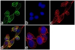
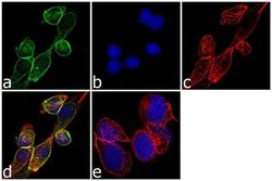
- Immunofluorescence analysis of GHRL was performed using 70% confluent log phase MKN-45 cells. The cells were fixed with 4% paraformaldehyde for 10 minutes, permeabilized with 0.1% Triton™ X-100 for 10 minutes, and blocked with 1% BSA for 1 hour at room temperature. The cells were labeled with Ghrelin Rabbit Polyclonal Antibody (Product # PA1-1070) at 2 µg/mL in 0.1% BSA and incubated for 3 hours at room temperature and then labeled with Goat anti-Rabbit IgG (H+L) Superclonal™ Secondary Antibody, Alexa Fluor® 488 conjugate (Product # A27034) a dilution of 1:2000 for 45 minutes at room temperature (Panel a: green). Nuclei (Panel b: blue) were stained with SlowFade® Gold Antifade Mountant with DAPI (Product # S36938). F-actin (Panel c: red) was stained with Alexa Fluor® 555 Rhodamine Phalloidin (Product # R415, 1:300). Panel d represents the merged image showing cytoplasmic localization. Panel e shows the no primary antibody control. The images were captured at 60X magnification.
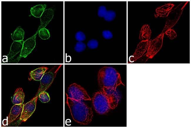
- Submitted by
- Invitrogen Antibodies (provider)
- Main image
- Experimental details
- Immunofluorescence analysis of GHRL was performed using 70% confluent log phase MKN-45 cells. The cells were fixed with 4% paraformaldehyde for 10 minutes, permeabilized with 0.1% Triton™ X-100 for 10 minutes, and blocked with 1% BSA for 1 hour at room temperature. The cells were labeled with Ghrelin Rabbit Polyclonal Antibody (Product # PA1-1070) at 2 µg/mL in 0.1% BSA and incubated for 3 hours at room temperature and then labeled with Goat anti-Rabbit IgG (Heavy Chain) Superclonal™ Secondary Antibody, Alexa Fluor® 488 conjugate (Product # A27034) a dilution of 1:2000 for 45 minutes at room temperature (Panel a: green). Nuclei (Panel b: blue) were stained with SlowFade® Gold Antifade Mountant with DAPI (Product # S36938). F-actin (Panel c: red) was stained with Alexa Fluor® 555 Rhodamine Phalloidin (Product # R415, 1:300). Panel d represents the merged image showing cytoplasmic localization. Panel e shows the no primary antibody control. The images were captured at 60X magnification.

Supportive validation
- Submitted by
- Invitrogen Antibodies (provider)
- Main image

- Experimental details
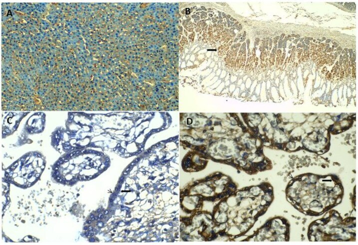
- Immunolocalization of LEAP-2 and Ghrelin protein in human tissues. ( A ). LEAP-2 protein is immunolocated in liver tissue (10x). ( B ). Ghrelin protein is immunolocated in human gastric mucosa (filled arrow in ( B ) (4x). ( C , D ) show immunostaining of LEAP-2 ( C ) and Ghrelin ( D ), respectively in cytotrophoblasts (filled arrow) (40x) and syncytiotrophoblasts (arrows) (40x) sections of human term placentas.