 Explore
 Validate
 Learn
Learn Western blot
Antibody data
- Antibody Data
- Antigen structure
- References [4]
- Comments [0]
- Validations
- Western blot [2]
- Immunohistochemistry [1]
Submit
Validation data
Reference
Comment
Report error
- Product number
- PAB12085 - Provider product page

- Provider
- Abnova Corporation
- Proper citation
- Abnova Corporation Cat#PAB12085, RRID:AB_1676267
- Product name
- OGG1 polyclonal antibody
- Antibody type
- Polyclonal
- Description
- Rabbit polyclonal antibody raised against synthetic peptide of OGG1.
- Storage
- Store at 4°C. Do not freeze.
Submitted references beta(2)-Adrenergic stimulation attenuates left ventricular remodeling, decreases apoptosis, and improves calcium homeostasis in a rodent model of ischemic cardiomyopathy.
The mitochondrial K-ATP channel opener, diazoxide, prevents ischemia-reperfusion injury in the rabbit spinal cord.
Protection of INS-1 cells from free fatty acid-induced apoptosis by targeting hOGG1 to mitochondria.
The p53 pathway promotes efficient mitochondrial DNA base excision repair in colorectal cancer cells.
Xydas S, Kherani AR, Chang JS, Klotz S, Hay I, Mutrie CJ, Moss GW, Gu A, Schulman AR, Gao D, Hu D, Wu EX, Wei C, Oz MC, Wang J
The Journal of pharmacology and experimental therapeutics 2006 May;317(2):553-61
The Journal of pharmacology and experimental therapeutics 2006 May;317(2):553-61
The mitochondrial K-ATP channel opener, diazoxide, prevents ischemia-reperfusion injury in the rabbit spinal cord.
Roseborough G, Gao D, Chen L, Trush MA, Zhou S, Williams GM, Wei C
The American journal of pathology 2006 May;168(5):1443-51
The American journal of pathology 2006 May;168(5):1443-51
Protection of INS-1 cells from free fatty acid-induced apoptosis by targeting hOGG1 to mitochondria.
Rachek LI, Thornley NP, Grishko VI, LeDoux SP, Wilson GL
Diabetes 2006 Apr;55(4):1022-8
Diabetes 2006 Apr;55(4):1022-8
The p53 pathway promotes efficient mitochondrial DNA base excision repair in colorectal cancer cells.
Chen D, Yu Z, Zhu Z, Lopez CD
Cancer research 2006 Apr 1;66(7):3485-94
Cancer research 2006 Apr 1;66(7):3485-94
No comments: Submit comment
Supportive validation
- Submitted by
- Abnova Corporation (provider)
- Main image
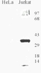
- Experimental details
- Western blot analysis of OGG1 in HeLa and Jurkat cell lysate with OGG1 polyclonal antibody (Cat # PAB12085).
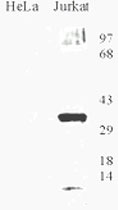
- Submitted by
- Abnova Corporation (provider)
- Main image

- Experimental details
- Western blot analysis of OGG1 of 1, 5, 10 ng titration of human recombinant Ogg1 protein with OGG1 polyclonal antibody (Cat # PAB12085).
Supportive validation
- Submitted by
- Abnova Corporation (provider)
- Main image
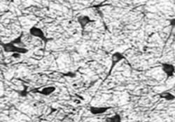
- Experimental details
- Immunohistochemical staining of OGG1 on formaldehyde fixed frozen sections of the substantia nigra from a Rhesus macaque with OGG1 polyclonal antibody (Cat # PAB12085). Photo courtesy of Glen Kisby, Oregon Health Sciences University. The tissue was a generous gift of Dr. Steven Kohama, Oregon National Primate Research Center.
- Validation comment
- Immunohistochemistry (Frozen sections)
