Explore
 Validate
 Learn
LearnPA5-40076
antibody from Invitrogen Antibodies
Targeting: RUNX1T1
AML1T1, CBFA2T1, CDR, ETO, MTG8, ZMYND2
 Western blot
Antibody data
- Antibody Data
- Antigen structure
- References [0]
- Comments [0]
- Validations
- Western blot [1]
- ELISA [2]
- Chromatin Immunoprecipitation [3]
- Other assay [2]
Submit
Validation data
Reference
Comment
Report error
- Product number
- PA5-40076 - Provider product page

- Provider
- Invitrogen Antibodies
- Product name
- RUNX1/RUNX1T1 Polyclonal Antibody
- Antibody type
- Polyclonal
- Antigen
- Synthetic peptide
- Reactivity
- Human
- Host
- Rabbit
- Isotype
- IgG
- Vial size
- 100 μL
- Concentration
- Conc. Not Determined
- Storage
- -20°C or -80°C if preferred
No comments: Submit comment
Supportive validation
- Submitted by
- Invitrogen Antibodies (provider)
- Main image
- Experimental details
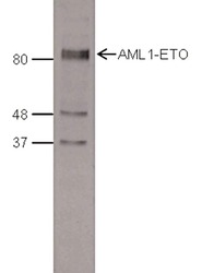
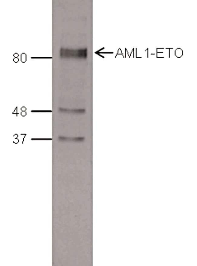
- Nuclear extracts of SKNO-1 cells (15 µg) were analyzed by Western Blot using a AML1-ETO polyclonal antibody (Product # PA5-40076) at a dilution of 1:1,000 in TBS-Tween containing 5% skimmed milk. The marker (kDa) is shown on the left, the position of the protein of interest is indicated on the right.
Supportive validation
- Submitted by
- Invitrogen Antibodies (provider)
- Main image

- Experimental details
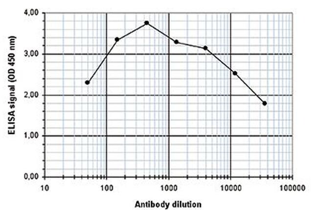
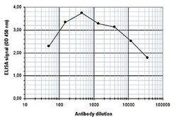
- To determine the titer of the antibody, an ELISA was performed using a serial dilution of the anti-AML1-ETO antibody (Product # PA5-40076). The plates were coated with the peptides used for immunization of the rabbit. By plotting the absorbance against the antibody dilution, the titer of the antibody was estimated to be 1:32,750.
- Submitted by
- Invitrogen Antibodies (provider)
- Main image
- Experimental details
- To determine the titer of the antibody, an ELISA was performed using a serial dilution of the anti-AML1-ETO antibody (Product # PA5-40076). The plates were coated with the peptides used for immunization of the rabbit. By plotting the absorbance against the antibody dilution, the titer of the antibody was estimated to be 1:32,750.

Supportive validation
- Submitted by
- Invitrogen Antibodies (provider)
- Main image
- Experimental details
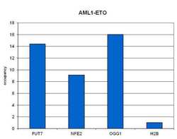
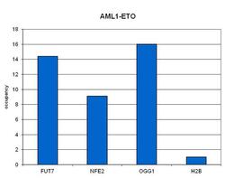
- ChIP assays were performed on Kasumi-1 cells using a AML1-ETO polyclonal antibody (Product # PA5-40076) and optimized primer pairs for qPCR. Sheared chromatin from 1.25 million cells and 4 µl of antibody were used per ChIP experiment. QPCR was performed on primers specific for the FUT7, NFE2 and OGG1 genes. Figure 1 shows the occupancy, calculated as the ratio + control/background for which the promoter of the H2B gene was used.

- Submitted by
- Invitrogen Antibodies (provider)
- Main image
- Experimental details
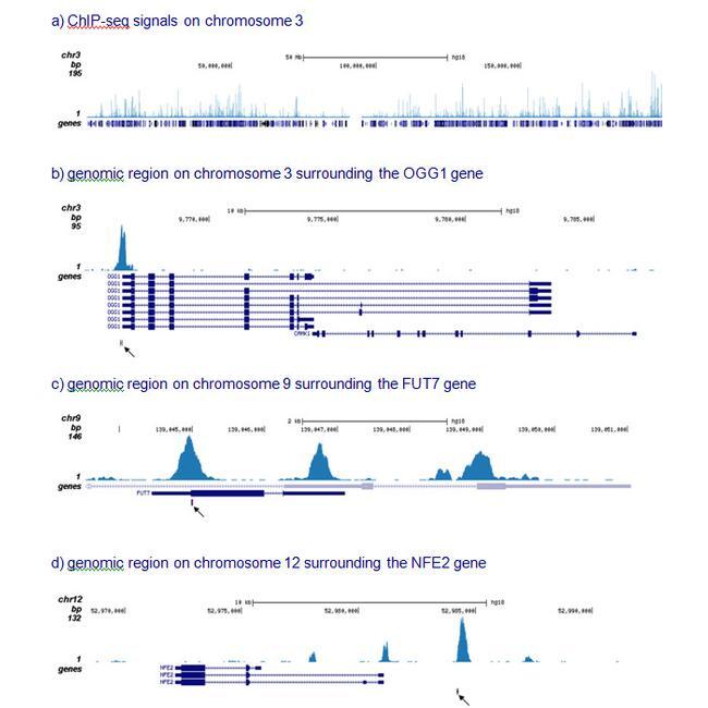
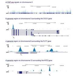
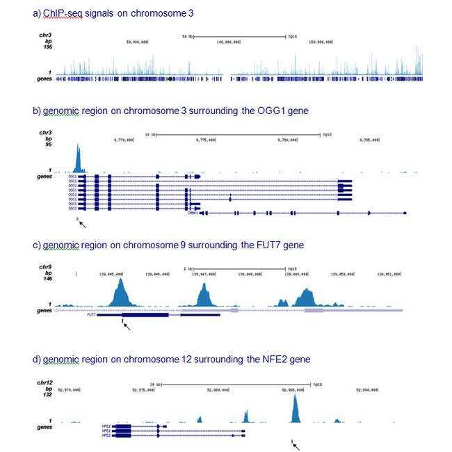
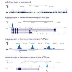
- ChIP assays were performed on Kasumi-1 cells using a AML1-ETO polyclonal antibody (Product # PA5-40076) and optimized primer pairs for qPCR. Sheared chromatin from 1.25 million cells and 4 µl of antibody were used per ChIP experiment. QPCR was performed on primers specific for the FUT7, NFE2 and OGG1 genes. The IP'd DNA of 6 ChIPs was pooled and analyzed with a Genome Analyzer. The 32 bp tags were aligned to the human reference genome (hg18) using the ELAND algorithm. Figure 2 shows the results of the complete chromosome 3 and three genomic regions surrounding the OGG1, FUT7 and NFE2 genes, respectively. The position of the PCR amplicon is indicated with an arrow.
- Submitted by
- Invitrogen Antibodies (provider)
- Main image
- Experimental details
- ChIP assays were performed on Kasumi-1 cells using a AML1-ETO polyclonal antibody (Product # PA5-40076) and optimized primer pairs for qPCR. Sheared chromatin from 1.25 million cells and 4 µl of antibody were used per ChIP experiment. QPCR was performed on primers specific for the FUT7, NFE2 and OGG1 genes. Figure 1 shows the occupancy, calculated as the ratio + control/background for which the promoter of the H2B gene was used.

Supportive validation
- Submitted by
- Invitrogen Antibodies (provider)
- Main image
- Experimental details
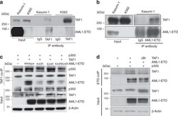
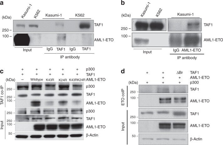
- Fig. 5 TAF1 associates with acetylated K43 on AE through its bromodomains. a TAF1 physically interacts with AE in Kasumi-1 cells. Co-immunoprecipitation was performed using anti-TAF1 antibody or normal mouse IgG. b Co-immunoprecipitation of TAF1 with AE using anti-ETO antibody or normal goat IgG. c Mutation of lysine-43 to arginine in AE reduces the interaction of AE with TAF1. 293T cells were transfected with p300, TAF1 and AE or AE mutants. Co-immunoprecipitation was performed using an anti-TAF1 antibody. d The deletion of the TAF1 bromodomain regions impairs its binding to AE. 293T cells were transfected with p300, AE, and TAF1 wild type or bromodomain deletion (DeltaBr) plasmids. Co-immunoprecipitation was performed using an anti-ETO antibody
- Submitted by
- Invitrogen Antibodies (provider)
- Main image
- Experimental details
- ChIP assays were performed on Kasumi-1 cells using a AML1-ETO polyclonal antibody (Product # PA5-40076) and optimized primer pairs for qPCR. Sheared chromatin from 1.25 million cells and 4 µl of antibody were used per ChIP experiment. QPCR was performed on primers specific for the FUT7, NFE2 and OGG1 genes. The IP'd DNA of 6 ChIPs was pooled and analyzed with a Genome Analyzer. The 32 bp tags were aligned to the human reference genome (hg18) using the ELAND algorithm. Figure 2 shows the results of the complete chromosome 3 and three genomic regions surrounding the OGG1, FUT7 and NFE2 genes, respectively. The position of the PCR amplicon is indicated with an arrow.