Explore
 Validate
 Learn
Learn Western blot
Antibody data
- Antibody Data
- Antigen structure
- References [0]
- Comments [0]
- Validations
- Western blot [1]
- Immunocytochemistry [1]
- Other assay [1]
Submit
Validation data
Reference
Comment
Report error
- Product number
- AF5410 - Provider product page

- Provider
- R&D Systems
- Product name
- Human/Mouse/Rat Cyclophilin B Antibody
- Antibody type
- Polyclonal
- Description
- Antigen Affinity-purified. Detects human, mouse, and rat Cyclophilin B in Western blots. In Western blots and direct ELISAs, less than 1% cross-reactivity with recombinant human Cyclophilin A is observed.
- Reactivity
- Human, Mouse, Rat
- Host
- Goat
- Conjugate
- Unconjugated
- Antigen sequence
P23284- Isotype
- IgG
- Vial size
- 100 ug
- Concentration
- LYOPH
- Storage
- Use a manual defrost freezer and avoid repeated freeze-thaw cycles. 12 months from date of receipt, -20 to -70 °C as supplied. 1 month, 2 to 8 °C under sterile conditions after reconstitution. 6 months, -20 to -70 °C under sterile conditions after reconstitution.
No comments: Submit comment
Supportive validation
- Submitted by
- R&D Systems (provider)
- Main image

- Experimental details
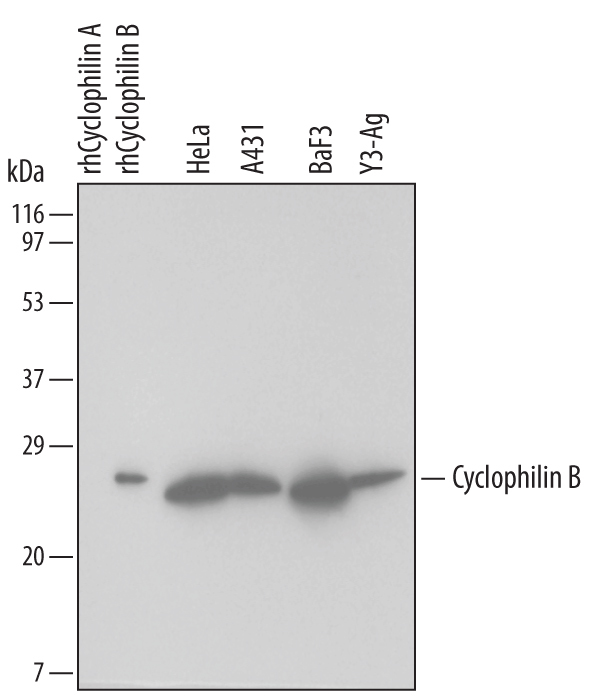
- Detection of Human/Mouse/Rat Cyclophilin B by Western Blot. Western blot shows lysates of HeLa human cervical epithelial carcinoma cell line, A431 human epithelial carcinoma cell line, BaF3 mouse pro-B cell line, and Y3-Ag rat myeloid cell line. PVDF membrane was probed with 1 µg/mL Goat Anti-Human/Mouse/Rat Cyclophilin B Antigen Affinity-purified Polyclonal Antibody (Catalog # AF5410) followed by HRP-conjugated Anti-Goat IgG Secondary Antibody (Catalog # HAF019). For additional reference, recombinant human Cyclophilin A and Cyclophilin B (5 ng/lane) were included. A specific band for Cyclophilin B was detected at approximately 24 kDa (as indicated). This experiment was conducted under reducing conditions and using Immunoblot Buffer Group 1.
Supportive validation
- Submitted by
- R&D Systems (provider)
- Main image
- Experimental details
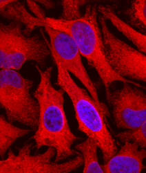
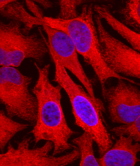
- Cyclophilin B in HeLa Human Cell Line. Cyclophilin B was detected in immersion fixed HeLa human cervical epithelial carcinoma cell line using Goat Anti-Human/Mouse/Rat Cyclophilin B Antigen Affinity-purified Polyclonal Antibody (Catalog # AF5410) at 10 µg/mL for 3 hours at room temperature. Cells were stained using the Northern-Lights™ 557-conjugated Anti-Goat IgG Secondary Antibody (red; Catalog # NL001) and counterstained with DAPI (blue). Specific staining was localized to cytoplasm. View our protocol for Fluorescent ICC Staining of Cells on Coverslips.
Supportive validation
- Submitted by
- R&D Systems (provider)
- Main image
- Experimental details
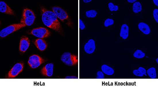
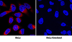
- Cyclophilin B in HeLa Human Cell Line. Cyclophilin B was detected in immersion fixed wildtype (left panel) but is not detected in Cyclophilin B knockout (right panel) HeLa human cervical epithelial carcinoma cell line using Goat Anti-Human/Mouse/Rat Cyclophilin B Antigen Affinity-purified Polyclonal Antibody (Catalog # AF5410) at 1 µg/mL for 3 hours at room temperature. Cells were stained using the NorthernLights™ 557-conjugated Anti-Goat IgG Secondary Antibody (red; Catalog # NL001) and counterstained with DAPI (blue). Specific staining was localized to cytoplasm. View our protocol for Fluorescent ICC Staining of Cells on Coverslips.